Biomedical Engineering Reference
In-Depth Information
Received
electric
signal
Imposed
electric
signal
Imposed
electric
signal
Chitosan
Protein
Analyte
Gold wire
Electrodeposition
Electrochemical
conjugation
Assembly
Transduction
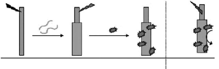
Figure 8.7
Gold wires are biofunctionalized using cathodic signals to electrodeposit chitosan and anodic signals to activate
the chitosan for protein assembly. (From Meyer, W. et al. 2009.
Biomacromolecules
10: 858-864. With permission.)
Figure 8.7, Meyer et al. use chitosan to serve as the interface between the protein-based
recognition element and a metal wire. Importantly, chitosan allows proteins to be assem-
bled in response to imposed electrical signals without the need for reactive reagents or
harsh conditions. Thus, biofunctionalization is simple, safe, and rapid. Further, the chito-
san coating is permeable to small molecules and allows the detection of electrochemically
active compounds that are either present in the solution or generated during the biological
recognition event. Thus, chitosan-coated electrodes can transduce chemical and biological
information into convenient electrical signals [59]. Yi et al. shows a sequence of steps in
which chitosan is first electrodeposited onto the patterned surface of our “chip,” the depos-
ited chitosan is next activated by GA, and then an amine-terminated single-stranded DNA
probe (20 bases) is conjugated onto the activated chitosan film. This chitosan-bound probe
DNA can hybridize with a fluorescently labeled target nucleic acid that has a complemen-
tary sequence [60].
8.3.7 electrospinning Method
Electrospinning is a progressive method that produces fibers ranging from the submicron
level to several nanometers in diameter in a high-voltage electrostatic field. As the electric
field surpasses a threshold value where the electrostatic repulsion force of surface charges
overcome surface tension, the charged fluid jet is ejected from the tip of the Taylor cone
and the bending jet produces highly stretched polymeric fiber with simultaneous rapid
evaporation of the solvent. Important parameters in electrospinning are not only polymer
and solution properties such as molecular weight, viscosity, conductivity, and surface ten-
sion, but also electrospinning conditions such as applied electric voltage, tip-to-collector
distance, feeding rate, and so on [61]. Nowadays, chitosan nanofibers can be promising
materials for many biomedical applications such as tissue templates, medical prostheses,
artificial organ, wound dressing, drug delivery, and pharmaceutical composition [62].
The electrospinning process was employed by Xu and coworkers to prepare a stabilized
chitosan nanofibrous membrane as a support for enzyme immobilization.
Figure 8.8
shows
a schematic representation of lipase immobilization on chitosan nanofibers. A chitosan
nanofibrous membrane was directly fabricated from a mixture solution of chitosan and
PVA and then treated with an NaOH solution in order to remove PVA and stabilize
the morphologies of the chitosan nanofibrous membrane in aqueous media. Lipase from
C. rugosa
was chosen as a model enzyme and immobilized on the prepared nanofibrous
Search WWH ::

Custom Search