Biomedical Engineering Reference
In-Depth Information
(a)
100
(b)
200
CPT lactone
CPT lactone
Before incubation
6h post incubation in PBS
Before incubation
6h post incubation in PBS
80
150
60
100
40
CPT carboxylate
50
20
CPT carboxylate
0
0
0
3
6
9
12
0
3
6
9
12
Time (min)
Time (min)
(c)
120
Free CPT
CPT-HGC
100
80
60
40
20
0
0
5
10
15
20
25
Time (h)
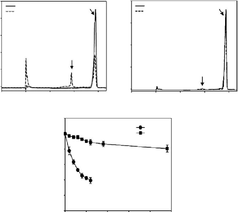
Figure 3.29
Protection effect of CPT-10 wt%-HGC nanoparticles on the lactone ring of CPT against hydrolysis over time
under physiological conditions (pH 7.4, 37°C). Reversed-phase HPLC chromatograms of (a) CPT and (b) CPT-
10 wt%-HGC nanoparticles before and after incubation for 6 h under physiological conditions (PBS, pH 7.4,
37°C). (b) Kinetic valuation of the rat of lactone ring opening for free CPT and CPT-10 wt%-HGC nanoparticles
evaluated by reversed-phase HPLC under physiological conditions (PBS, pH 7.4, 37°C).
5.5-labeled CPT-HGC nanoparticles with 10 mg/kg of CPT, the time-dependent excretion
profile, tumor accumulation, and tissue distribution of Cy 5.5-labeled CPT-HGC nanopar-
ticles in tumor-bearing mice were evaluated using the Explore Optix system and the
Kodak Image Station 4000 MM. First, the time-dependent excretion profile of CPT-HGC
nanoparticles was clearly visualized by monitoring real-time NIR fluorescence signals in
the whole body
(
Figure 3.30a).
After the i.v. injection of Cy 5.5-labeled CPT-HGC nanopar-
ticles, the NIR fluorescence intensity immediately increased in the whole body, due to the
rapid circulation of Cy 5.5-labeled CPT-HGC nanoparticles. However, the NIR fluores-
cence signal in the whole body decreased as time elapsed, which was indicative of excre-
tion by renal clearance. It is deduced that nano-sized drug carriers in the blood might be
dissociated and biodegraded
in vivo
and then excreted by renal clearance. Importantly,
CPT-HGC nanoparticles displayed strong fluorescence signals in tumor regions, com-
pared to the whole body (Figure 3.30b and c). Furthermore,
ex vivo
fluorescence images
Search WWH ::

Custom Search