Biomedical Engineering Reference
In-Depth Information
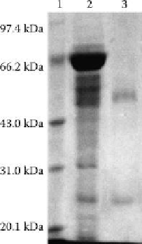
Figure 3.3
SDS-PAGE gel stained by Coomassie Blue. Lane 1, marker; lane 2, 4% of the plasma before adsorption; lane 3,
supernatant after adsorption of chitosan.
is reached when there is not too much interaction of platelets with the material surface.
Therefore, platelet adhesion on microspheres from blood is an important test for the
evaluation of blood compatibility of microspheres. Platelet adhesion and activation are
two important steps that regulate the formation of the thrombus and medical device
rejection. During the initial stage of surface activation, the change in conformation of
adsorbed proteins exposes RGD sequences that are sensitive to the platelet GPIIb/III
receptor. When platelets are surface activated, they progress through a sequence of mor-
phological changes. The surface activation contributes to the change in the organization
of the cytoskeleton, and thus increases the surface area of platelets by the formation of
pseudopods. The platelets thus, adhered and activated, go through a sequence of cytoskel-
etal events and increase in endoplasmic Ca
2+
concentration, polymerization of action fila-
ments, thrombin activation, and release of cytoskeletal granule contents, as well as
platelet aggregation. The extent of shape change and spread area has been related to the
surface energy of polymer materials [87]. Chitosan microspheres make them activated.
Chou et al. [88] demonstrated that chitosan is an effective inducer of rabbit platelet adhe-
sion and aggregation, and explained that the mechanisms of action of chitosan may be
associated with the increase of Ca
2+
mobilization and the enhancement of expression of
the GPIIb-IIIa complex. It has also been reported that chitosan and chitin enhance plate-
let aggregation due to their amino residues [89]. In acidic solution, the amine groups of
chitosan are protonated to −NH
3
+
, which makes chitosan cationic in nature, allowing for
electrostatic interactions with the negatively charged biological molecules on the platelet
surface [90].
The shortening of clotting time by chitosan may be related to not only platelet aggregation
but also erythrocyte aggregation [89]. Earlier it was observed that the procoagulation proper-
ties of chitosan were partly due to erythrocytes [91]. Chitosan may induce the adhesion of
erythrocytes with its amino groups or by forming a three-dimensional network structure in
blood that captures the erythrocytes and then makes them aggregates [92]. In this system
[93], chitosan microspheres were directly immersed into erythrocyte suspension without
any blood proteins. The surface of chitosan microspheres greatly enhanced erythrocyte
a g g l u t i n at i o n o r a g g l o m e r at i o n , w h i c h i n d i c at e d t h at c h it o s a n c a n d i r e c t l y i n du c e e r y t h r o c y t e

Search WWH ::

Custom Search