Biomedical Engineering Reference
In-Depth Information
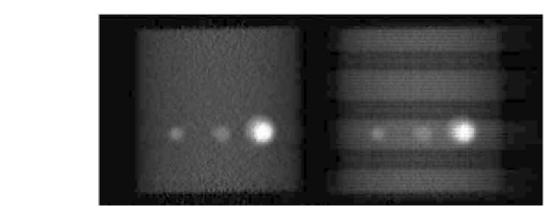
FIGURE 4.6: Phantom with active spheres (coronal slice), reconstructed
using normalization file (left) and without correction for normalization effects
(right).
no normalization for the image reconstruction is used, these gaps are clearly
visible in the reconstructed PET images (Figure 4.6).
4.1.5
Dead
time
eects
(loss
of
count
rate)
(PET
and
SPECT)
Dead time is the time a detection system needs after the detection of
one event to be prepared for the detection of another event. This means, if
another event occurs in the dead time this new event is ignored. Instead of
not detecting the second event, for energy-sensitive detectors the signal of the
first event and the second event may overlap and lead to wrong signals. Such
merged events may fall outside the energy window of the detection electronics
and hence be lost for the image reconstruction. Such events are called dead
time losses. The number of dead time losses gets smaller as the dead time
of the detection system is shorter. Naturally, higher photon flux leads to a
higher probability of another photon not being registered. This means that
the measured photons are not proportional to the real photon flux at higher
count rates. The fractional dead time of a system is defined as:
Measured Count Rate
Count Rate with Ideal Linear Behaving System
:
Dt =
(4.4)
4.1.6 Partial volume effects (PET and SPECT)
Due to the limited spatial resolution of SPECT and PET tomographs,
partial volume effects occur when the acquired objects are smaller than a
resolution-volume element of the machine. Partial volume effects lead to false
measured activity concentration, either too high or too low depending on


Search WWH ::

Custom Search