Biomedical Engineering Reference
In-Depth Information
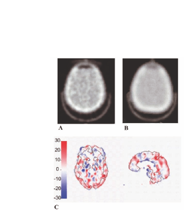
FIGURE11.4: MR-AC for brain PET: atlas-based attenuation correction.
(A) Attenuation map measured through a PET transmission scan; (B) Attenu-
ation map obtained through atlas registration and addition of the head holder;
(C) Coronal and saggital view of voxel-by-voxel relative differences between
PET attenuation corrected using attenuation maps A and B. (From [33].)


Search WWH ::

Custom Search