Biomedical Engineering Reference
In-Depth Information
(a)ExactPerfusion(b)Reconstructed
Perfusion
(c)ExactSpillover (d)Reconstructed
Spillover
(e)ExactSequence,Frame8(f)StandardEM,Frame8(g)ReconstructedSequence,
Frame8
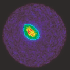
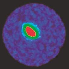
FIGURE9.3: A combined reconstruction/parameter identification process
of a synthetic myocardial perfusion example. Figure 9.3(a) and Figure 9.3(b)
show the exact perfusion and the reconstruction with the method proposed
in [3]. Figure 9.3(c) and Figure 9.3(d) show the exact arterial spillover and its
reconstruction. Figure 9.3(e){Figure 9.3(g) show the 8th frame of the underly-
ing exact image sequence, the frame-independent standard EM reconstruction
and the reconstruction method proposed in [3].
(a)
(b)
(c)
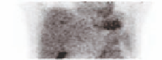
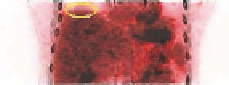
FIGURE9.5: Representation of one temporal bin of the acquisition gated
with the respiration: (a) gated PET image non corrected for attenuation, (b)
gated CT image, (c) overlaid image of both PET and CT gated images. A
misalignment of 6/7mm between the PET and CT images can be seen at the
level of the diaphragm on the overlaid image inside the yellow circle.


Search WWH ::

Custom Search