Biomedical Engineering Reference
In-Depth Information
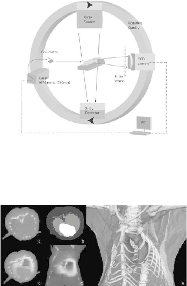
FIGURE 12.10: Combined FMT-XCT setup.
properties, structural priors based on the anatomy are used to guide the re-
construction and the XCT images are used for combined 3D visualization of
the fluorescence signal together with anatomy [1, 22]. Figure 12.11 shows re-
sults obtained with a hybrid FMT-XCT system. The anatomical segmentation
FIGURE 12.11: (See color insert.) Reconstruction based on data from
combined FMT-XCT setup. (a) X-ray slice. (b) Segmentation of X-ray data
in lungs, heart, bone and remaining tissue. (c) Reconstruction of fluorescent
biodistribution in lung, transversal slice and (d) sagittal slice. (e) 3D-hybrid
visualization. (From [22].)


Search WWH ::

Custom Search