Biomedical Engineering Reference
In-Depth Information
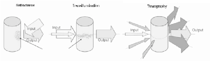
FIGURE 12.2: In fluorescence reflectance imaging, excitation light is ex-
panded on the object surface and fluorescence light is collected from the same
side of the object. In transillumination mode, illumination and detection are
performed on opposite sides. In fluorescence tomography, the object is illumi-
nated from different angles in transillumination mode. (From [34].)
fluorochromes. The light emitted by the fluorochromes is then captured by
a camera using appropriate filters. An alternative method to epi-illuminant
imaging is transillumination imaging, based on the same principle, but with
source and detectors placed on opposite sides of the tissue. An advantage of
transillumination is the larger feasible penetration depth. Due to the nonlinear
dependencies of light propagation through tissue, significant uncertainty on
the exact depth of the recorded signal exists with both methods. The depth of
the signal can be more accurately resolved when tomographic imaging is used.
In this case transillumination images from multiple source-detector configu-
rations are recorded and combined to a three-dimensional reconstruction of
the internal fluorochrome distribution. This technique is termed fluorescence
molecular tomography (FMT) and is the main subject of this chapter.
The principle of operation in FMT resembles that of X-ray computed to-
mography (CT) in that tissue is illuminated from different angles and at dif-
ferent positions and a mathematical formulation is used to describe photon
propagation in tissue. However, a major difference between optical tomogra-
phy and tomographic methods based on high-energy rays is that photons in
the optical range are highly scattered by tissue organelles and membranes.
Photons do not propagate in straight lines when traveling through tissue, but
become diffuse within a few millimeters of propagation. The diffusive nature
of the light propagation through tissue limits the quantification ability and
maximum resolution that can be achieved. Therefore, FMT is mainly con-
cerned with the localization and quantification of bulk signals from specific
fluorescent entities indicating cellular and molecular activity.
One of the most recent technological evolutions in the field of FMT has
been the development of multimodality systems, in which FMT is combined
with X-ray CT or MRI. A straightforward benefit of hybrid methods is that
they allow the seamless co-registration of the images obtained, since all the
modalities employed visualize the object of interest under identical placement.


Search WWH ::

Custom Search