Biomedical Engineering Reference
In-Depth Information
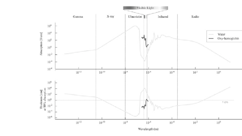
FIGURE 1.2: (See color insert.) The electromagnetic spectrum showing
the absorption coecient in water at different wavelengths. Both, gamma
radiation and visible light have low attenuation in water; oxy-hemoglobin
adds a considerable attenuation (compiled from [1, 2, 3]).
acquisition, data processing, image generation, image processing, and image
analysis (Figure 1.3).
Since acquired data are always affected by physical limitations of the ac-
quisition hardware, corrections have to be applied at a very early stage in
order to improve data and correct for these effects. In PET and SPECT imag-
ing, this involves correction for dead time, detector eciencies, etc. In optical
imaging light scattering in biological tissue is the main limitation for accurate
reconstruction of planar or even tomographic images. Highly sophisticated
reconstruction schemes and correction have to be applied to generate semi-
quantitative images. Many of these corrections need to be applied at the level
of data processing.
After data acquisition, the pre-corrected data are usually reconstructed by
imaging reconstruction techniques aiming for generating either planar images
or a three-dimensional tomographic volume. Since this is the most important
processing step that defines and determines the quality of the final results,
most of the corrections are directly integrated into the image reconstruction
procedure. For PET (and partly for SPECT), corrections for scattered pho-
tons, decay, and attenuation are usually peformed during image reconstruc-
tion.
Finally, the reconstructed images may be post-processed to further enhance
images and improve quantitative accuracy. Noise reduction by image filtering,


Search WWH ::

Custom Search