Biomedical Engineering Reference
In-Depth Information
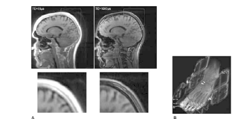
FIGURE 11.10: MR images acquired with a 3D ultrashort echo (UTE)
sequences. (A) Sagittal brain section acquired with short echo: 0.07 ms (left)
and late echo: 1 ms (right). (B) Angled view of a human foot. Note, bone
yields a high signal and so do parts of the coil housing.
lesions and body parts with high bone contents, need to show whether ignoring
bone is a clinically viable option.
Not accounting for bone in MR-based PET-AC is not an option for quan-
titative brain imaging: Receptor studies for example require an accuracy of
less than 5% [7].
11.4.2 MR imaging with ultrashort echo time (UTE)
Instead of performing advanced image segmentation methods on standard
MR images one may utilize dedicated MR sequences, such as ultrashort echo
time (UTE) sequences [39, 32] that yield signal even from cortical bone (Figure
11.10). Typically, the use of just a single UTE image does not enable one to
distinguish bone from non-bone tissues. However, when combined with a late
echo image it is, in principle, possible to detect bone as the structure that
yields a signal on the short echo image, but not on the late echo image (Figure
11.10A-B). By using multi-echo sequences [11] the 2 images can be acquired
in one scan. While it seems promising for brain applications, UTE may not
be acceptable as part of whole-body imaging protocols since acquisition time
is on the order of several minutes per bed position.


Search WWH ::

Custom Search