Biomedical Engineering Reference
In-Depth Information
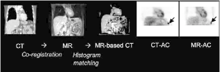
Single-station, transverse T1-weighted VIBE MR images were used to gen-
erate pseudo-CT images. First, the MR images were co-registered to the CT
images using non-linear, curvature-regularized co-registration in conjunction
with mutual information. Second, the MR voxel value intensity distribution
was matched to that of the co-registered CT image. MR-CT intensity trans-
formation was performed in a 3-step process based on a histogram-matching
algorithm. PET images were reconstructed on the PET/CT console following
attenuation correction based on CT transmission images (PET
CTAC
) and MR-
based, pseudo-CT images (PET
MRAC
); see Figure 11.6. The authors demon-
strated that histogram matching is a feasible technique to transform MR to
pseudo-CT attenuation values if the MR image quality is high. The study il-
lustrated the need for accurate patient positioning between the MR and PET,
but did not further quantify such effects.
Martinez-Moller et al. [23] proposed an approach to MR-AC where the
attenuation map is segmented into background, lungs, fat, and soft tissue,
which can be clearly delineated on MRI. The authors then evaluated the effect
of \ignoring" bone tissue. Their study included 35 patients who had received
18
F-FDG PET/CT. On 52 lesions they used a CT-derived attenuation map
that was segmented into the above four tissue classes, resulting in average
SUV dierences of 8% 3% (mean SD) for n = 21 bone lesions, 4% 2%
for n = 16 neck lesions, and 2%3% for n = 15 lung lesions. The largest SUV
dierence was an underestimation of 13:1% for a lesion in the pelvic bone.
The authors then applied the Dixon segmentation method [9] as a proof of
concept that such a 4-class attenuation map can be derived from an MR image
(Figure 11.7). The Dixon segmentation was complemented by a component
analysis to detect the lungs, and a morphological closing filter to avoid clas-
FIGURE 11.6: MR-AC for torso applications [3]. From top to bottom: CT
images from PET/CT studies are co-registered to available MR. CT-MR his-
togram matching yields images with pseudo-CT attenuation values that are
used for MR-AC. PET images following CT-AC and MR-AC show severe dif-
ferences if the MR images inherit artifacts from suboptimal imaging protocols.
In case of good MR image quality (see thorax) and accurate co-registration
MR-AC based on histogram matching appears feasible.


Search WWH ::

Custom Search