Biomedical Engineering Reference
In-Depth Information
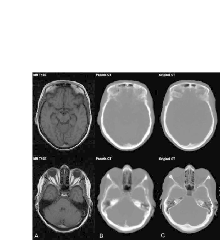
FIGURE 11.5: MR-AC for brain PET using the atlas-based method by
Hofmann et al. [16]. Axial slices through patient data with mid-plane sections
on top and lower brain sections on the bottom. (A) T1w spin-echo MR, (B)
pseudo-CT as predicted from the atlas-based MR-AC and (C) original CT
image. Note, the visual similarity between the pseudo-CT and the original
CT.


Search WWH ::

Custom Search