Biomedical Engineering Reference
In-Depth Information
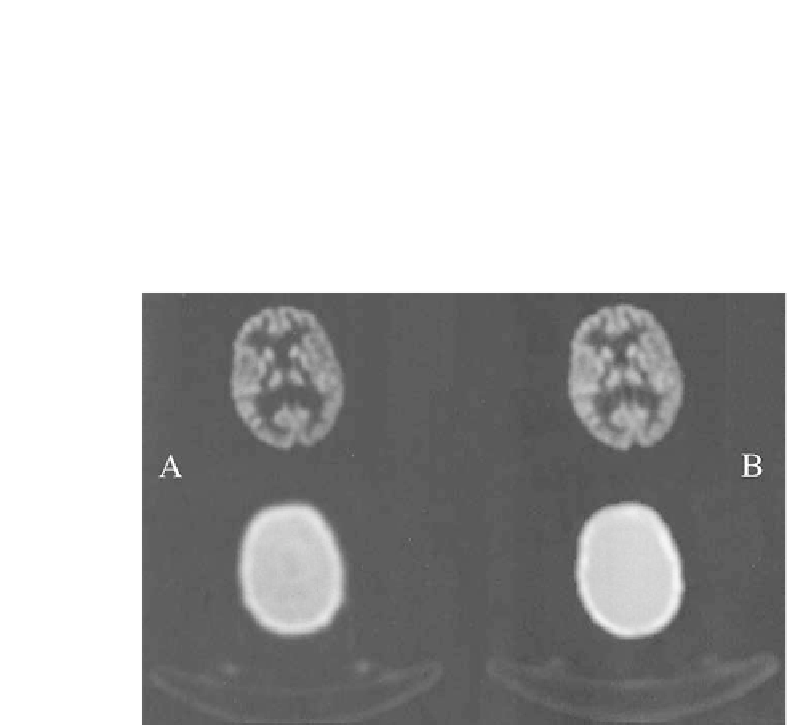
FIGURE 11.2: MR-AC for brain PET [41]. Axial slices through FDG-PET
of the cerebellum of a patient following standard TX-AC (A) and MR-AC (B)
with PET on top and the corresponding attenuation maps on the bottom.
PET images appear visually similar with a slightly better signal-to-noise ratio
following MR-AC.


Search WWH ::

Custom Search