Biomedical Engineering Reference
In-Depth Information
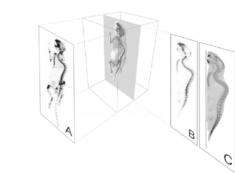
FIGURE 1.1: (See color insert.) Two different biomedical imaging princi-
ples demonstrated on a
18
18F-fluorid PET bone scan of a mouse: (A) projection
image showing the maximum intensity projection; (B) single PET slice out of
the 3D tomographic volume; (C) same slice but fused with CT data.
with co-registered anatomical information, thus combining the best of the two
imaging worlds.
Two different imaging principles have been developed in emission tomog-
raphy: projection imaging and tomography.
Similar to the principle of a camera, a projection is showing a two-
dimension picture of the tracer activity projected onto a surface outside the
object. If our eyes were sensitive to gamma rays we could directly see the
emitted \light" shining through the human body (or animal) as a projec-
tion image (Figure 1.1 A). Volumes of high tracer accumulation would appear
bright, areas of low tracer uptake would barely be visible.
Tomography is an imaging technique where the biological object is shown
as a stack of two-dimensional slices. Slices through the object of interest are
visualized as if the observer would be looking directly onto these slices (Fig-
ure 1.1 B and C). In contrast to projection imaging, tomography is a three-
dimensional imaging method showing details of the local tracer distribution.


Search WWH ::

Custom Search