Biomedical Engineering Reference
In-Depth Information
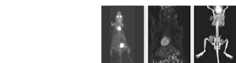
(a)
(b)
FIGURE 10.3: (See color insert.) PET-SPECT-CT. (a) Siemens Inveon
r
MultiModality. (b) Preclinical studies (courtesy of University of Wisconsin,
Madison, WI and Eberhard Karls University Tuebingen, Tuebingen, Ger-
many).
SPECT(/CT) and PET(/CT) systems and image fusion would then have to
be done during the processing step retrospectively.
10.3 The combination with MR
In the 1980s, magnetic resonance imaging (MRI) was introduced to clinical
practice. It quickly became a standard tool in the diagnosis of soft-tissue
abnormalities due to its high contrast and the ability to acquire easily oblique
slices. The absence of ionizing radiation also helped the acceptance of this new
modality.
Magnetic resonance uses the precession of particles with a spin and mag-
netic momentum in a magnetic field. A strong static magnetic field aligns these
particles. When applying RF pulses at the resonance frequency, the particles
will be pushed out of their stable condition absorbing the electromagnetic
energy, but will then return back to the baseline by emitting the absorbed
electromagnetic energy. The relaxation time and the amplitude of this sig-
nal is related to the chemical condition of the particle and its concentration.
Images are reconstructed based on either the longitudinal relaxation time T1
(related to magnetic moments of surrounding nuclei) or the transversal relax-
ation time T2 (determined by the frequency of the collisions of the molecules)
or a combination of both. The most common nucleus used for MR-imaging is
hydrogen, but other nuclei are used as well.
While general MR imaging provides superb images with high soft-tissue
contrast, more sophisticated pulse sequences and imaging techniques allow,
for example, flow and perfusion images, determination of spatial distribu-


Search WWH ::

Custom Search