Biomedical Engineering Reference
In-Depth Information
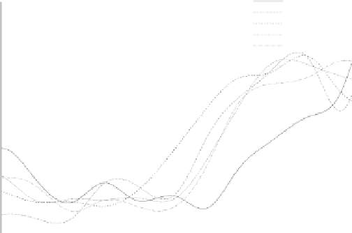
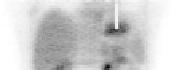
(a)
(b)
(c)
(d)
(e)
non−corrected
reference gated Frame
elastic corrected image using the CT series
elastic corrected image using the AC PET series
elastic corrected image using the non−AC PET series
Voxels position
(f)
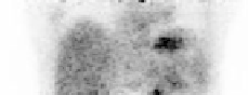
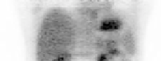
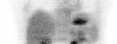
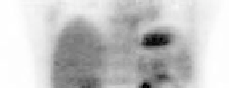
FIGURE 9.6: Comparison of (a) the gated image chosen as reference for
the motion compensation reconstruction and (b) the non-corrected image,
with the three motion-compensated images reconstructed using the elastic
transformation parameters derived using (c) the 4D CT images, (d) the 4D
PET images reconstructed with attenuation correction and (e) the 4D PET
images reconstructed without attenuation correction. For the three motion-
compensated images, all the PET data acquired at different time bins are elas-
tically realigned into the specific time point corresponding to this reference-
gated frame. The attenuation of the PET emission data is performed using
the same 4D CT images as used for image c. As shown in graph (f), visual
differences in terms of position can be clearly observed at the level of the
diaphragm, below the heart.


Search WWH ::

Custom Search