Biomedical Engineering Reference
In-Depth Information
(a) Exact Perfusion
(b) Reconstructed
Perfusion
(c) Exact Spillover
(d) Reconstructed
Spillover
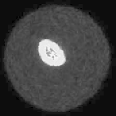
(e) Exact Sequence, Frame 8
(f) Standard EM, Frame 8
(g) Reconstructed Sequence,
Frame 8
FIGURE 9.3: (See color insert.) A combined reconstruction/parameter
identification process of a synthetic myocardial perfusion example. Figure
9.3(a) and Figure 9.3(b) show the exact perfusion and the reconstruction with
the method proposed in [3]. Figure 9.3(c) and Figure 9.3(d) show the exact
arterial spillover and its reconstruction. Figures 9.3(e){(g) show the 8th frame
of the underlying exact image sequence, the frame-independent standard EM
reconstruction and the reconstruction method proposed in [3].
9.3 Combined reconstruction and motion correction
Algorithms developed in context of Section 9.2 have also been applied to
other applications where similar problems appear; e.g., Grotus et al [18] used
a general 4D method to reconstruct respiratory-gated PET data. Addition-
ally, next to the application of known methods on these new type problems,
an independent theory has been investigated by several authors and will be
presented in this section.
One of the parameters affecting quantitation in emission tomography (ET)
imaging of the thoracic and abdominal regions is respiratory motion. Respi-
ratory motion has been shown to reduce the accuracy of determining func-
tional lesion volumes and associated recovered activity concentrations [41, 8]


Search WWH ::

Custom Search