Biomedical Engineering Reference
In-Depth Information
2
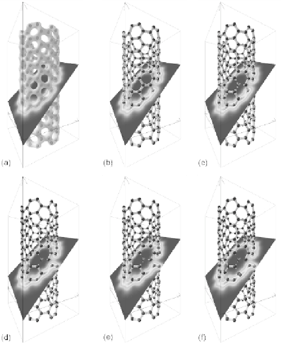
we can see that all of the bonds in the structure are sp
bonds (as
expected), since all bonds present in Figs. 7.7a and 7.8a also appear
as dumbbell-shaped surfaces between the atoms. In Figs. 7.7c-f and
8c-f 1(P), 2(P), 3(P), and 3(R) hydrogen atom are adsorbed, and the
changes in the sp
2
network associated with the H adsorption are
evident. Note that C-H bonds are not shown.
Figure 7.7
The ECD in a plane bisecting the pristine (6, 6) CNT with (a) the
total bonding iso-surface, (b) the sp
2
iso-surface, along with (c)
the sp
2
iso-surface with one H atom adsorbed, (d) with two H
atoms adsorbed, (e) with three H atoms adsorbed in the 3(P)
configuration, and (f) with three H atoms adsorbed in the 3(R)
configuration.