Biomedical Engineering Reference
In-Depth Information
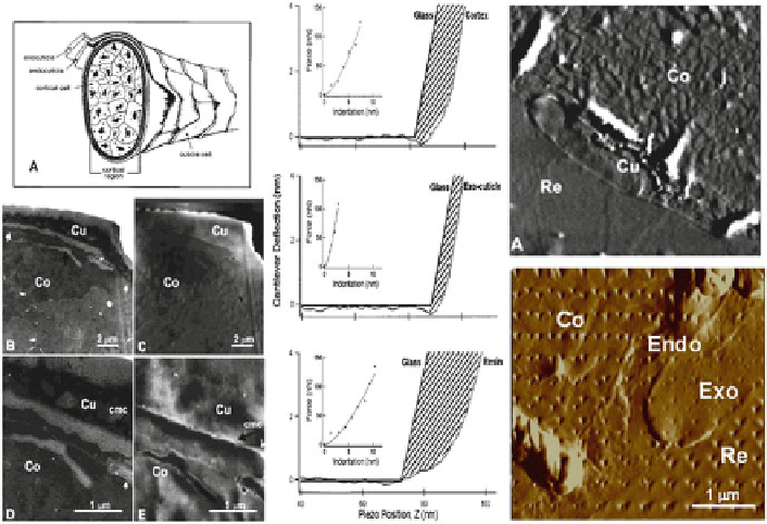
Fig. 11
Left
: Schematic diagram of wool fi ber (
a
). TEM image [(
b
), higher magnifi cation in (
d
)]
and AFM image [(
c
), higher magnifi cation in (
e
)] of same wool fi ber region. Cu, cuticle cells and
some portion of the cortical region (Co). AFM and TEM images correlate well with respect to
identifi ed subcellular structures.
Center
: Force curves on cortex (
top
), exocuticle (
middle
), and
embedding resin (
bottom
).
Solid line
is collected on hard glass surface.
Larger shaded area
indi-
cates more elastic surface.
Top right
image shows corresponding AFM image; the cortex (Co),
cuticle (Cu), and embedding resin (Re).
Bottom right
image shows surface after indentation with
diamond tip to create specifi c patterns on wool fi ber; exocuticle (Exo), endocuticle (Endo), and
cortex (Co). For details, see Parbhu et al. (
1999
)
complete biocomposite (Weiner and Wagner
1998
). Degradation of this nonfi brillar
organic matrix (Braidotti et al.
1997
), for instance by heat treatment, leads to an
alteration of the failure mechanism from delamination of the bone along the con-
tours of the trabeculae to a more rugged, nondirectional and diffuse crack propaga-
tion (Fantner et al.
2004
). Similar degradation of the organic matrix leading to brittle
bones may be involved in the increased brittleness of older or osteoporotic bone.
Using AFM, the mechanics of bone and the importance of the organic matrix can be
studied at the nanoscale. Both scanning electron microscopy (SEM) and AFM stud-
ies show that mineralized collagen fi brils are interconnected by a glue-like organic
matrix that hold the fi brils together ( Fig.
12
, left panel) (Fantner et al.
2005
) and are
stretched out upon separation of the collagen fi brils.
To test the mechanical strength of the organic matrix material, pieces of bone
were attached to both the AFM tip and cantilever, and force spectroscopy resulted
in pulling curves that show the fi laments breaking in a step-like manner (Fig.
12
,
middle and right panels) (Fantner et al.
2005
). Furthermore, from the force curves,
the energy dissipation during separation of bone fi brils can be calculated. Since
AFM is performed in buffer solution, the effect of ion concentration can also be