Biomedical Engineering Reference
In-Depth Information
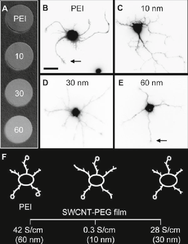
Fig. 3
Image of 12 mm in diameter glass coverslips (
a
) coated, from top to bottom, with: PEI
(standard), 10-, 30-, and 60-nm-thick SWCNT-PEG fi lms. Fluorescence images of live hippocam-
pal neurons grown on coverslips coated with PEI coated (
b
) or SWCNT-PEG fi lms of varying
thickness/conductivity (
c
, 10-nm thick;
d
, 30-nm thick;
e
, 60-nm thick). Arrows indicate growth
cones. (
b-e
) Scale bar, 20 m m. (
f
) Drawing summarizing the effects of the conductivity of SWCNT
substrates on neurite outgrowth, growth cones, and body size. The neurite outgrowth was signifi -
cantly greater in neurons grown on the 10-nm thick SWCNT-PEG fi lms. The average area of the
neuron cell body grown on the 30-nm thick SWCNT fi lms was enlarged. Neurons grown on the
PEI had a signifi cantly higher number of growth cones than those grown on 10- and 30-nm SWCNT
fi lms, but not higher than those grown on 60-nm SWCNT fi lms. Conductivity of each fi lm is listed
in S/cm; PEI is not conductive. Modifi ed from (Malarkey et al.
2009
)