Biomedical Engineering Reference
In-Depth Information
impact on the quality, interpretation, and relevance of biological or physiological
results obtained using quantum dot-labeling nanotechnologies.
5
Labeling Reactive Gliosis in Retinal Tissue Sections
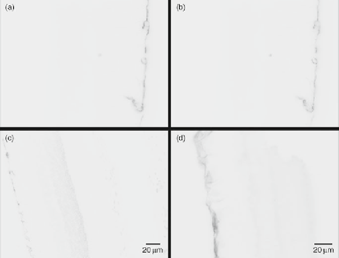
In the normal neural retina, GFAP expression is associated with the astrocyte layer in
the inner nuclear layer and the endfeet of Müller glial cells near the retinal capillar-
ies. Quantum dot labeling of GFAP in control sections of rat retina showed that only
Müller cell endfeet and astrocytes were GFAP positive, with no GFAP upregulation
and no nonspecifi c binding (Fig.
6a, b
). The high specifi city and signal-to-noise ratio
Fig. 6
Control labeling of the noninjured rat neural sensory retina for GFAP. (
a
,
b
) Anti-GFAP
antibody functionalized quantum dot conjugates specifi cally label only Muller cell endfeet-associ-
ated retinal capillaries and astrocytes associated in the inner nuclear layer associated with retinal
ganglion cells. Two slices from a wide-fi eld nonconfocal image stack are shown, and display no
observable nonspecifi c labeling despite the use of nonconfocal mode. (
c
,
d
) Wide-fi eld nonconfocal
standard ICC using an anti-40,6-diamidino-2-phenylindole(GFAP)-conjugated primary antibody
and FITC fl uorophore-tagged secondary antibody. A nonspecifi c nuclear DAPI stain was used to
visualize the other retinal layers. Note the more diffuse labeling using FITC compared to the quan-
tum dots and the presence of some nonspecifi c labeling in the distal layers of the retina. Panels (
a
),
(
b
), and (
d
) were taken at 40× and 50-ms exposure times while panel (
c
) was taken at 20× at a 50-ms
exposure. All micrographs are 10-mm slices. Reproduced from Pathak et al. (
2009
)