Biomedical Engineering Reference
In-Depth Information
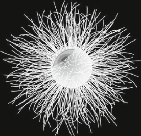
Fig. 1
Cartoon structure of a
typical functionalized
quantum dot. The heavy metal
core is shielded from the
biological environment by an
outer shell. The outer shell is
in turn chemically
functionalized with
biologically relevant
molecules, such as antibodies
and other peptides (
black
), for
specifi c binding to target
epitopes, for example, on cells
can be chemically functionalized to target proteins at high ligand-receptor densi-
ties. Recent work has shown that, at least in some cellular systems, quantum dots
conjugated with natural ligands are readily internalized into cells, do not interfere
with intracellular signaling, and are nontoxic.
For neuroscience, quantum dots represent a tool of signifi cant potential.
Besides offering an alternative to traditional ICC, they are particularly valuable
for studies of neurons and glia. Quantum dots can be used to visualize, measure,
and track individual molecular events using fl uorescence microscopy; they pro-
vide the ability to visualize and track dynamic molecular processes over extended
periods (e.g., from seconds to many minutes). These properties are diffi cult to
achieve using other techniques or approaches. For example, quantum dots are use-
ful for experiments that are limited by the restricted anatomy of neuronal and glial
interactions, such as the small size of the synaptic cleft, or between an astrocyte
process and a neuron. Because of their extremely small size and optical resolu-
tion, they are also well-suited for tracking the molecular dynamics of intracellular
and/or intercellular molecular processes over long timescales. However, it should
be appreciated that the hydrodynamic radius of functionalized quantum dots is
larger (15-20 nm) than their actual size of 5-8 nm. Recent studies using quantum
dots in neuroscience illustrate the potential of this technology. Triller and col-
leagues used antibody functionalized quantum dots to track the lateral diffusion of
glycine receptors in cultures of primary spinal cord neurons (Dahan et al.
2003
) .
They were able to track the trajectory of individual glycine receptors for tens of
minutes at spatial resolutions of 5-10 nm, demonstrating that the diffusion dynam-
ics varied depending on whether the receptors were synaptic, perisynaptic, or
extrasynaptic. Vu, Desai, and colleagues tagged nerve growth factor (bNGF) to