Biomedical Engineering Reference
In-Depth Information
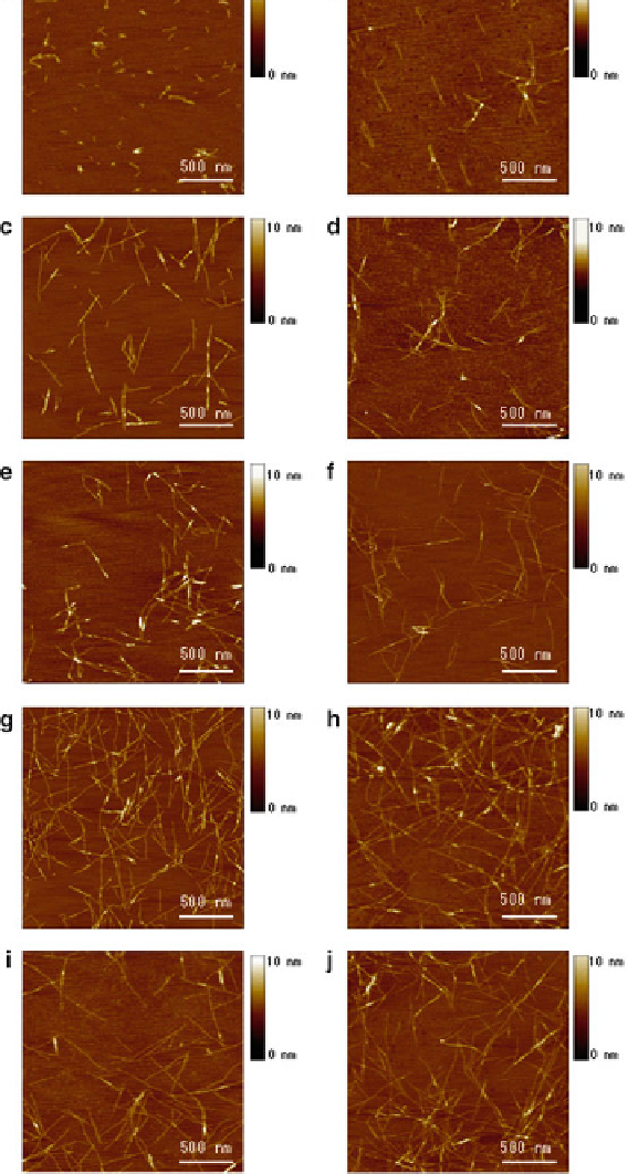
Fig. 10
AFM images of RADA16-I nanofi bers at various time points after sonication. The obser-
vations were made using AFM immediately after sample preparation. (
a)
1 min; (
b
) 2 min; (
c
) 4 min;
(
d
) 8 min; (
e
) 16 min; (
f
) 32 min; (
g
) 64 min; (
h
) 2 h; (
i
) 4 h; (
j
) 24 h after sonication. Note the
elongation and reassembly of the peptide nanofi bers over time. By ~1-2 h, these self-assembling
peptide nanofi bers have nearly fully reassembled