Biomedical Engineering Reference
In-Depth Information
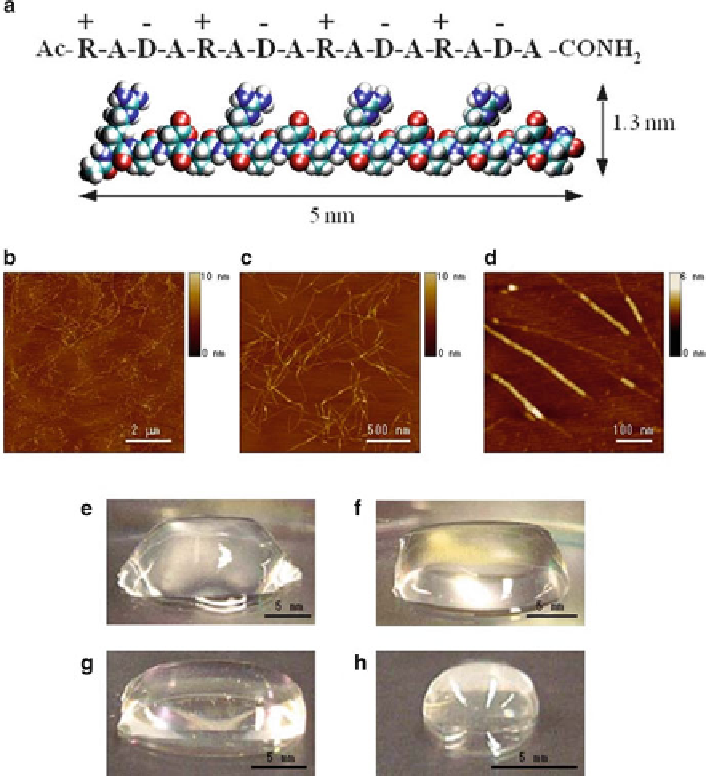
Fig. 9
Peptide RADA16-I. (
a
) Amino acid sequence and molecular model of RADA16-I (~5 nm
long, 1.3 nm wide and 0.8 nm thick). (
b-d
) AFM images of RADA16-I nanofi ber scaffold. Image
size, (
b
) 8 × 8 m m, (
c)
2 × 2 mm, and (
d)
0.5 × 0.5 mm. Note variability in height by ~1.3 nm of dif-
ferent segments within individual nanofi ber suggesting its double-layered structure (
d
). (
e-h
)
Photographs of RADA16-I hydrogel at various condition, (
e
) 0.5 wt.% (pH 7.5), (
f
) 0.1 wt.% (pH
7.5, Tris.HCl), (
g
) 0.1 wt.% (pH 7.5, PBS) before sonication, (
h
) reassembled RADA16-I hydrogel
after four times of sonication, respectively
as distinct increase in height of about 1.3-1.5 nm can be seen; this height corre-
sponds to the addition of a single thickness of a peptide. Figure
9e
-h shows the
peptide scaffold hydrogel formation at various concentrations [0.6-3 mM, or
1-5 mg/ml (w/v)] and water content (99.5-99.9%) (Yokoi et al.
2005
) . The scaffold
hydrogel is completely transparent, which is a very important requirement for accu-
rate image collections for uses in 3-D tissue cell cultures.