Biomedical Engineering Reference
In-Depth Information
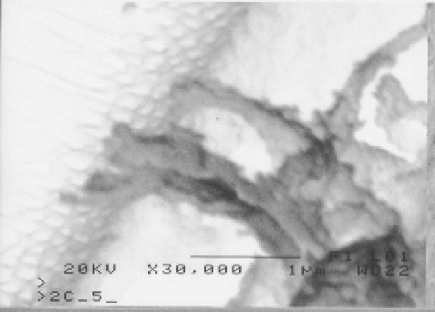
Fig. 2
An osteoblast on a nanoporous alumina membrane extends processes into the pores
removed. This process creates a periodic concave pattern on the surface. These
concavities act as the nucleation sites for the pores that are created in the second anod-
ization and etching step. The second anodization is a repeat of the fi rst anodization to
deposit an even layer of alumina on the surface of the sheet. After this anodization, the
protective polymer layer is stripped from the backside to expose the aluminum and the
sheet is soaked in a mixture of copper chloride and hydrochloric acid. The etching
removes the exposed aluminum and exposes the alumina layer beneath. The fi nal step
is to remove the alumina layer deposited in the second anodization process. This is
done by placing a few drops of 10% phosphoric acid on the alumina surface and
allowing it to soak until the pores are exposed. The SEM image of a nonporous alu-
mina scaffold produced using the above outline process is shown in Fig.
1b
.
Human fetal osteoblasts were cultured on the nanoporous membranes and their
adhesion, proliferation, performance and ECM production were monitored. These
alumina nanoporous scaffolds were found to increase osteoblast adhesion over non-
porous membranes. Adhesion and proliferation were increased for up to 4 days of
culture (Popat et al.
2005
) while matrix production was increased for 4 weeks of
culture. Further modifi cation of the porous membranes was achieved by immobiliz-
ing RGDC moieties onto the alumina (Swan et al.
2005b
). The addition of this pep-
tide sequence did not clog the pores and increased osteoblast adhesion as well as
matrix production 2 days into culture. The introduction of nanopores changes what
the cells sense as they grow on the surface. How individual cells are interacting with
the nanoporous membrane is of particular importance. Scanning electron micros-
copy of osteoblasts growing on the nanoporous membranes shows that cells extend
processes into the nanopores (Fig.
2
). The cells appear to be exploring their environment