Biomedical Engineering Reference
In-Depth Information
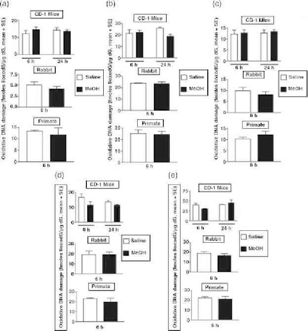
FIGURE 7.26 MeOH-initiated 8-oxo-2
0
deoxyguanine (8-oxodG) formation
in the lung, liver, kidney, bone marrow, and spleen of male CD-1 mice, NZW
rabbits, and cynomolgous monkeys in 6 hours. Animals were treated intra-
peritoneally with a single dose of 2.0 g/kg bw MeOH (20% [w/v] in sterile
saline) or saline vehicle (controls) and sacrificed at 6 or 24 hours post-
injection. Genomic DNA was isolated and analyzed for oxidatively damaged
DNA damage reflected by the formation of 8-oxodG in the (a) lung, (b) liver,
(c) kidney, (d) bone marrow, and (e) spleen of each species. Values are
mean
3 for rabbit and monkeys, respectively.
Source: Modified from McCallum et al. (2011a,b).
þ
SE; N
¼
4 for mice and N
¼
7.4.4.2 Hydroxynonenal-Histidine Protein Adducts On the basis of
our negative findings for MeOH-dependent ROS-mediated DNA dam-
age and conflicting reports in the literature of MeOH-induced lipid
peroxidation (Skrzydlewska et al., 2000; Parthasarathy et al., 2006a;