Biomedical Engineering Reference
In-Depth Information
colon, is divided into the cecum, ascending colon, transverse colon, descending colon,
sigmoid colon, rectum, and anus. The salivary glands, liver, gallbladder, and pancreas
deliver digestive secretions and help in digestion and absorption of drugs.
10.2.2.1 Stomach
Surface of the stomach is divided into three well-defined tissue layers: the muscle,
the submucosal and mucosal layers. Because of the smaller size, lack of villi, thick
mucosal layer, short residence time, and smaller absorption surface area, absorption
from the stomach is minimal. The major function of the gastric mucosal epithelium is
to secrete hydrochloric acid, pepsin, intrinsic factor, and bicarbonate
[29,30]
. Acidic
pH affects the folded structure of protein and causes protein denaturation. These
denatured proteins are more likely to be attacked by pepsin because of increased
exposure of the peptide bonds to pepsin, which breaks down the proteins into
peptides. However, the peptide bond breakage in the stomach is incomplete because
pepsin can only break peptide bonds between specific amino acid sequences in pep-
tides and proteins.
10.2.2.2 Small Intestine
The small intestine is a tubelike structure approximately 200 in. long with a radius of
approximately 0.75 in., which is subdivided into three anatomical regions: the duode-
num (the first 10 in.), the jejunum (the second 80 in.), and the ileum (the last 110 in.)
[29]
. Ingested food and macromolecules are enzymatically hydrolyzed to smaller
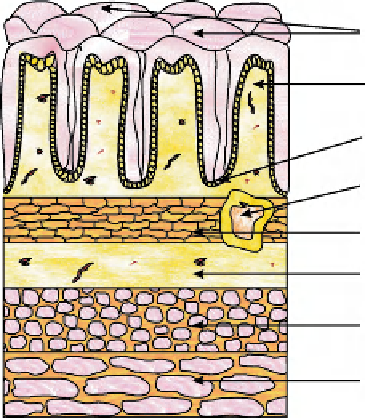
molecules in the small intestine. The wall of the intestinal membrane has four dif-
ferent layers: the mucosa, submucosa, muscularis, and serosa. These tissue layers
are depicted in
Fig. 10.1
. The innermost, or luminal, surface is the mucosal layer,
Figure 10.1
General
histology of the GI
tract.
Surface epithelium
Lamina propria
Crypt of lieberkuhn
Lymphoid
Muscularis mucosae
Submucosa
Circular muscle of
muscularis externa
Longitudinal muscle of
muscularis externa
Search WWH ::

Custom Search