Biology Reference
In-Depth Information
2001). This chapter briefly reports unpublished observations on the axial mobility of an adult
captive, male
Inia
that was housed at the Pittsburgh Zoo (Figure 2), and examines the
mobility of vertebral regions in this mammal based on measurements from video recorded
images. This dolphin was previously studied by Fish (1997) and Buchholtz (2001). Herein,
movements from the dorsal perspective are examined in the first attempt to quantify the
degree of lateral axial flexibility of this species. Anatomical attributes of river dolphins
generally, and
Inia geoffrensis
in particular, are reviewed in the context of our findings.
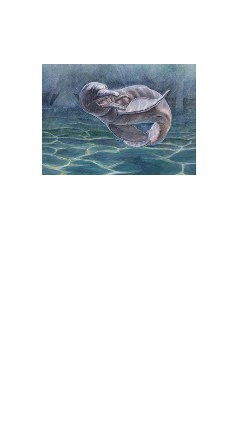
Figure 2. Illustration of a captive (Pittsburgh Zoo) Amazon River dolphin (
Inia geoffrensis
) pursuing
live fish (© 2008, Timothy D. Smith). This image emphasizes the extreme mobility of the species,
afforded by unfused cervical vertebrae, relatively long vertebral centra throughout the torso, and large
paddle-shaped pectoral flippers.
O
BSERVATIONS OF A
C
APTIVE
R
IVER
D
OLPHIN
:
M
EASUREMENT
T
ECHNIQUES
Despite some chronic health problems (Bonar & Wagner, 2003), the male Amazon River
dolphin at the Pittsburgh Zoo lived for more than three decades. This dolphin was fed a diet
of live brown trout and had daily interactions with trainers or zoo staff. On four separate days,
mobility at the head and neck (atlantooccipital joint; cervical vertebrae) and torso (primarily
intervertebral joints of lumbar and caudal vertebrae) was examined by videotaping the
dolphin during training or feeding sessions. These sessions were taped simultaneously from
lateral and dorsal perspectives. In order to synchronize the two videotapes for analysis, a
trainer initiated the session by targeting the dolphin or dropping a fish into the tank. The
lateral videotapes were used to determine when the body axis of the animal was roughly
parallel to the floor of the tank, thus ensuring little distortion on measurements taken from the
dorsal view. The dorsal videotapes were then used to obtain measurements of body mobility,
but only when a parallel position had been confirmed based on synchronized portions of the
lateral videotape. These particular moments were captured and saved as bitmap files for
analysis using Scion Image software (NIH).
Search WWH ::

Custom Search