Image Processing Reference
In-Depth Information
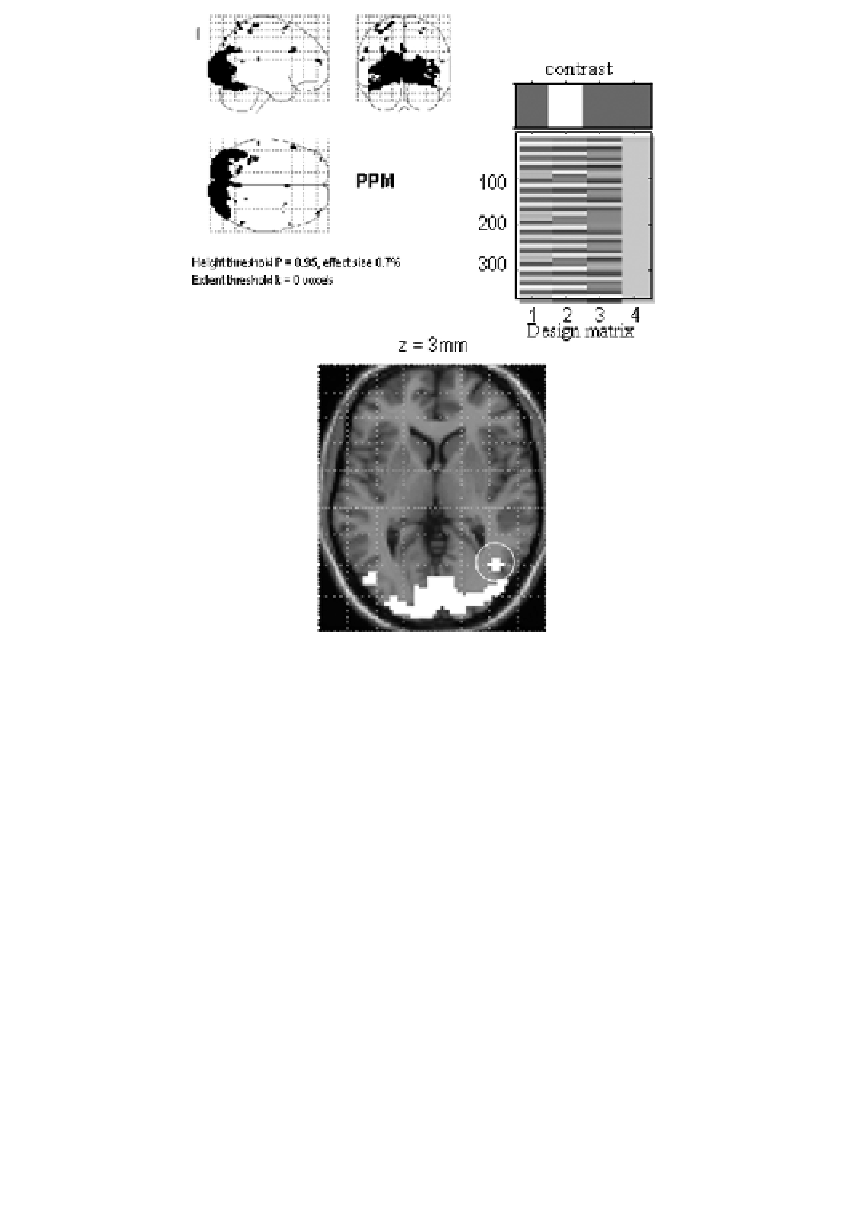
FIGURE 17.4
PPM for the fMRI study of attention to visual motion. The display format
in the lower panel uses an axial slice through extrastriate regions, but the thresholds are
the same as employed in maximum intensity projections (upper panels). The activation
threshold for the PPM was 0.7. As can be imputed from the design matrix, the statistical
model of evoked responses comprised of boxcar regressors convolved with a canonical
hemodynamic response function.
using a conventional SPM procedure and the empirical Bayesian approach
described in the previous section. The ensuing SPMs and PPMs are presented in
Figure 17.4 and
Figure 17.5
. We used a contrast that tested for the effect of visual
motion above and beyond that due to photic stimulation with stationary dots.
The difference between the PPM and SPM is immediately apparent on inspec-
tion of Figure 17.4 and Figure 17.5. Here, the threshold for the PPM was 0.7%
(equivalent to percentage whole brain mean signal). Only voxels that exceed 95%
confidence are shown. These are restricted to visual and extrastriate cortex
involved in motion processing. The critical thing to note is that the corresponding
SPM identifies a smaller number of voxels than the PPM. Indeed, the SPM appears
to have missed a critical and bilaterally represented part of the V5 complex


Search WWH ::

Custom Search