Image Processing Reference
In-Depth Information
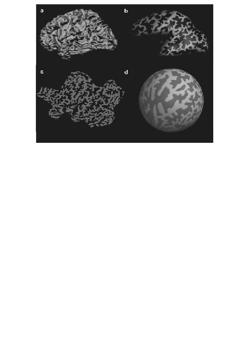
FIGURE 15.7
Morphing of the cortical surface. (a) Folded, (b) inflated, (c) flattened, and
(d) sphered
representation of the cortex.
representation of the 3-D pattern of cortical activation without loss of the lobular
structure of the telencephalon (see, e.g.,
Reference 30
and
Reference 32
). Inflated
representations can be further processed to obtain flattened representations of the
cortex, which are used in the study of retinotopy [49-52] and tonotopy [53], or
to obtain “spherical” representations of the cortex, which are used in advanced
approaches of cortex-based normalization and realignment of the brains of dif-
ferent subjects [54-56] (Figure 15.7).
REFERENCES
1.
Ogawa, S., Tank, D., Menon, R., Ellermann, J.M., Kim, S.G., Merkle, H., and
Ugurbil, K. (1992). Intrinsic signal changes accompanying sensory stimulation:
functional brain mapping using MRI.
Proc. Natl. Acad. Sci.
USA
89: 5951-5955.
2.
Kwong, K.K., Belliveau, J.W., Chesler, D.A., Goldberg, I.E., Weisskoff, R.M.,
Poncelet, B.P., Kennedy, D.N., Hoppel, B.E., Cohen, M.S., Turner, R., Cheng,
H.M., Brady T.J., and Rosen, B.R. (1992). Dynamic magnetic resonance imaging
of human brain activity during primary sensory stimulation.
Proc. Natl. Acad. Sci.
USA
89: 5675-5679.
3.
Bandettini, P.A., Wong, E.C., Hinks, R.S., Tikofsky, R.S., and Hyde, J.S. (1992).
Time course EPI of human brain function during task activation.
Magn. Reson.
Med.
25: 390-398.


Search WWH ::

Custom Search