Image Processing Reference
In-Depth Information
A
B
−
100
−
50
0
+50
+100
1
0
20
30
40
Displacement (
µ
m)
Shear Stiffness (kPa)
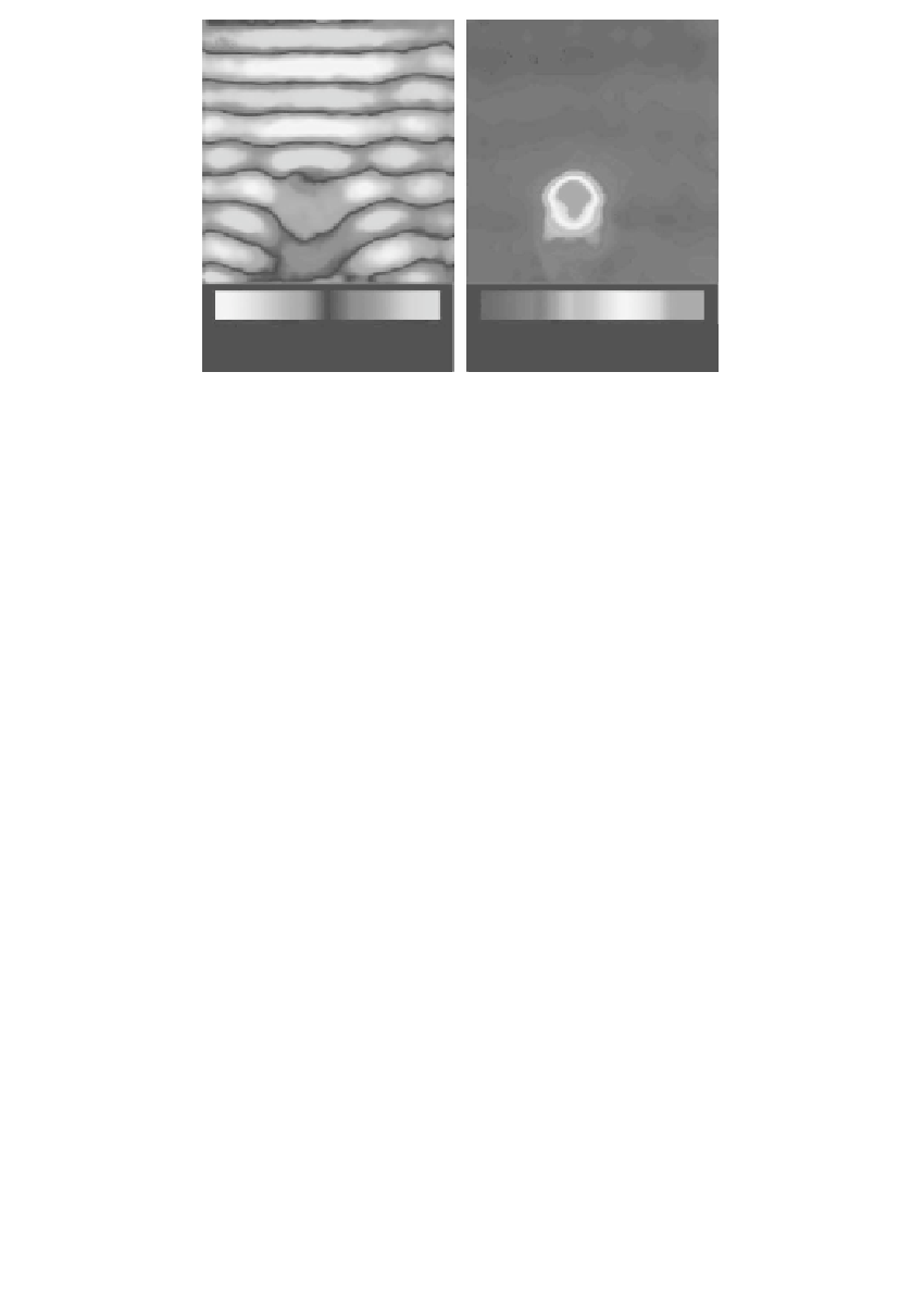
FIGURE 14.2
(a) Shear waves propagating in a phantom with an embedded 1.5-cm-
diameter cylinder of stiffer gel. Shear waves at 300 Hz were applied at the top margin
of the gel block, with transverse motion oriented orthogonal to the plane of the image.
(b) The elastogram based on LFE processing clearly depicts the object even though it is
significantly smaller than the wavelength in the stiff material.
sequences are also in common use but require longer acquisition times (several
minutes to tens of minutes).
The phase images reflect the displacement of spins due to acoustic strain
wave propagation in the medium and are termed
. Such an image of
propagating acoustic waves in a tissue-simulating agarose gel phantom is shown
in Figure 14.2a. The wave propagation depends on the elasticity of the material
at each location in the object, so inversion of the data can yield elastograms as
in Figure 14.2b. Experiments to assess the sensitivity of the shear-wave-imaging
method at low amplitudes of mechanical excitation demonstrated that shear waves
with displacements of less than 100 nm can be readily observed [8].
Mechanical excitation can be provided by a moving-coil driver with the
imager providing the static magnetic field. Trigger pulses are provided by the
sequencing computer of the MR imager and are fed to a function generator that
produces a waveform that is amplified and applied to the coil of the actuator.
Transverse stresses applied to a flat contact plate provide planar shear waves.
Piezoelectric drivers can also be used for generating shear waves. Mechanical
excitation can also be applied longitudinally, with mode conversion leading to
shear waves being generated inside the object [33,34]. Focused ultrasound also
can be used to generate shear waves that can be imaged with MRE [35]. The
ultrasound beam is temporally modulated to create cyclic variation in acoustic
radiation pressure at the focus of the ultrasound source, which can be located
deep within an object.
By adjusting the phase offset between the mechanical excitation and the
wave images
oscillating magnetic gradient (
θ
in
Figure 14.1
), acoustic-wave images can be


Search WWH ::

Custom Search