Image Processing Reference
In-Depth Information
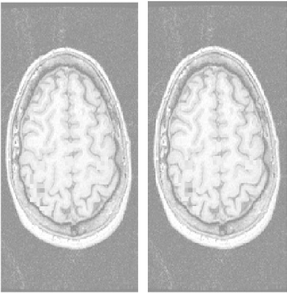
FIGURE 7.8
fMRI activation maps related to (left) not-registered and (right) registered
data.
hand with the thumb. In this experiment, the motor area of the cervical cortex is
activated in a well-known way. The fMRI data set is composed of 60 temporal
acquisitions (acquired every 3 sec), 128
∞
128
∞
24 images each, with a pixel size
of 2.2
2.2 mm and a slice thickness of 7 mm. A standard multislice EPI sequence
was used. Functional images are mapped on a structural data set, composed of 8
slices, 256
∞
1.1 mm and a slice thickness
of 7 mm. The registration operation was performed using the AFNI software, based
on the AIR approach for the implementation of the registration.
Figure 7.8 shows the activation maps related to not-registered (left) and reg-
istered (right) data. It is clear from the figure that the registration is not able to
correct all the activation artifacts, because of the strong correlation between subject
movement and the imposed paradigm. However, registered images show a reduc-
tion of the artifacts in brain borders as well as an improved delineation of the
∞
256 pixels each, with a pixel size of 1.1
∞
activation zone.
Figure 7.9
shows the values of the six roto-translation parameters
used to perform the registration along the image sequence. The detected move-
ments are in general very small (less than 2 mm). Continuous movements during
the examination are effectively revealed, while subcutaneous movements generated
by paradigm changes are often missed.


Search WWH ::

Custom Search