Biology Reference
In-Depth Information
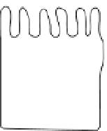
(A)
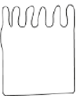
(B)
Apical
ES
ESp
Apical
compartment
Sertoli cell
eSp
Apical
junctional
complex
Epithelial cell
TJ
rSp
AJ
Sy
DS
Nu
Basal
ES
Nu
BTB
BTB
GJ
Nu
Nu
Basal
compartment
Sy
Sg
Basal lamina
Hemidesmosome
Focal adhesion
complex
Basement membrane
Figure 6.1
Differences in the morphological layouts of junction types between a typical
epithelium/endothelium and the seminiferous epithelium.
(A) For the junctional complex in
typical epithelia/endothelia, TJs, which are responsible for sealing the intercellular space to
create the barrier function by regulating paracellular and transcellular transport, are located
at the apical region of the lateral membrane between adjacent epithelial/endothelial cells.
Underneath TJs, there are AJs that contribute to most of the adhesive force of the apical
junctional complex by connecting to a dense F-actin network, creating the zonula adherens
plaque, to be followed by desmosomes. Both TJ and AJ are actin-based cell-cell anchoring
junctions, whereas DS is intermediate filament-based cell-cell anchoring junction. Other
junctional molecules such as GJs, which are not part of the junctional complex, are localized
basal to the junctional complex (constituted by TJ, AJ and DS). (B) Unlike the junctional com-
plex in typical epithelia which are furthest away from the basal lamina, the BTB in seminifer-
ous epithelium is located near the basement membrane (a modified form of extracellular
matrix in the testis). Instead of being arranged as discrete structure as in other epithelia/
endothelia, TJs, basal ES (a testis-specific actin-rich AJ) and GJs are
coexisting
at the BTB,
which together with DS are all involved in creating the BTB. The BTB physically separates the
seminiferous epithelium into the basal and apical (adluminal) compartments. Spermatogo-
nia and preleptotene spermatocytes reside at the basal compartment, and preleptotene
spermatocytes that arise at stage VII-VIII of the epithelial cycle in the rat testis are the only
germ cells that can traverse the BTB. After traversing the BTB, spermatocytes undergo meio-
sis and eventually differentiate into elongating/elongated spermatids, and spermatids (step
8-19 spermatids in the rat testis) anchored to the Sertoli cells by apical ES. Furthermore,
hemidesmosomes (intermediate filament-based cell-matrix anchoring junction) and focal
adhesion complexes (FAC, or known as focal contacts, an actin-based cell-matrix anchoring
junction) are also found in most epithelia, but FAC is absent in the seminiferous epithelium.
Abbreviations used: Sg, spermatogonium; Sy, spermatocyte; rSp, round spermatid; eSp,
elongating spermatid; ESp, elongated spermatid; Nu, Sertoli cell nucleus; DS, desmosome;
AJ, adherens junction; GJ, gap junction; TJ, tight junction; ES, ectoplasmic specialization. For
color version of this igure, the reader is referred to the online version of this topic.
During the transit of preleptotene spermatocytes conneced in “clones”
via intercellular bridges from the basal to the apical compartment, sper-
matocytes have first to travel across a blood-tissue junctional barrier, which
physically separates the two compartments (
Fig. 6.1
). This junctional bar-
rier, which located near the basement membrane, is formed by adjacent


Search WWH ::

Custom Search