Biology Reference
In-Depth Information
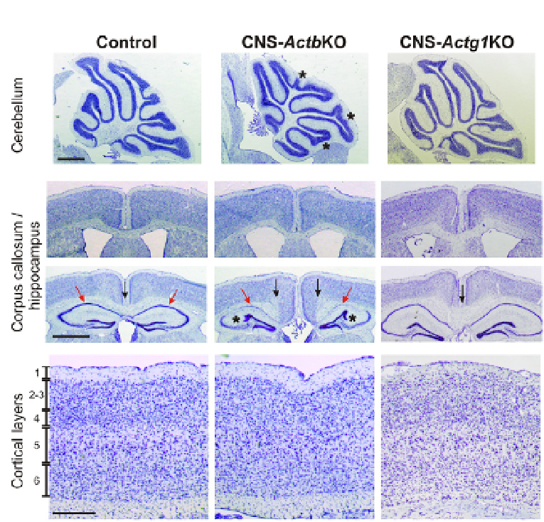
Figure 4.4
Histological phenotypes following ablation of β- but not γ-actin in the brain.
Midsagittal sections through the cerebellum of CNS-
Actb
KO mice revealed abnormali-
ties in the positioning and morphology of the cerebellar folia (asterisks) that was not
observed in CNS-
Actg1
KO animals. Scale bar 0.5 mm. Coronal sections through the ros-
tral portion of the brain (second panel from top) showed the normal crossing of the
rostral corpus callosum in all genotypes. However, a localized defect in corpus callosum
crossing was observed in CNS-
Actb
KO mice in more caudal sections overlying the hip-
pocampus (black arrows in third from top panel). An abnormal invagination of the den-
tate gyrus (asterisks) and decreased CA1 staining (red arrows) was also only apparent
in CNS-
Actb
KO animals. Scale bar 1 mm. Sagittal sections through the cortex (bottom
panel) revealed no significant defects in cortical layering in either CNS-
Actb
or
Actg1
KO
brain sections. Scale bar 0.3 mm. Control and CNS-
Actb
KO images for comparison from
Cheever et al. (2012).
For interpretation of the references to color in this figure legend,
the reader is referred to the online version of this topic.
(
Dugina et al., 2009
). Cells depleted of β-actin by RNAi-mediated knock-
down or genetic ablation both exhibit impaired growth and significantly
elevated numbers of multinucleate cells while γ-actin-deficient cells do
not (
Bunnell et al., 2011
;
Dugina et al., 2009
), further supporting the idea
that β-actin is specifically involved in cellular constriction events. Although
Search WWH ::

Custom Search