Biomedical Engineering Reference
In-Depth Information
Case 11
Nevus of Ota
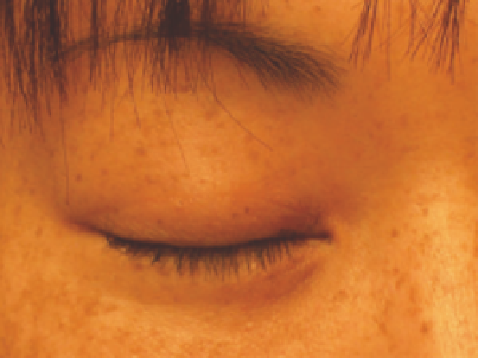
This 26-year-old woman is concerned about the pigmented lesion above and below the left eyelid (Fig. 3.11A). Because of the
close proximity of the lesion to the eye, a stainless steel, internal eye shield was inserted into the eye prior to treatment. The
patient required someone to drive her to and from the offi ce in case of any blurry vision from the use of the topical anesthetic
and lubrication required for inserting the eye shield. Local anesthesia was performed with 1% lidocaine with epinephrine
infi ltrated into the lesion prior to each procedure to minimize pain. Figure 3.11B shows the result after one treatment with the
QS alexandrite laser at 9 J/cm
2
; there is 75% resolution of the lesion. Figure 3.11C shows the result 6 months after a second
treatment with the QS ruby laser at 5 J/cm
2
: there is complete resolution of the lesion.
(
A
)
(
B
)
(
C
)
Figure 3.11
(
A
) Pigmented lesion above and below the left eyelid. (
B
) The result after one treatment with a Q-switched alexandrite laser at 9 J/cm
2
; there is 75%
resolution of the lesion. (
C
) 6 months after a second treatment with the Q-switched ruby laser at 5 J/cm
2
.
Source
: Photos courtesy of Mitchel P. Goldman, MD.
Case 12
Nevus of Ito
This 3-year-old presented with a dark brown linear nevus on the lateral aspect of the neck extending to the left chin (Fig. 3.12A).
Figure 3.12(B) shows the appearance 6 years after 3-monthly treatments with a QS ruby laser at 5 J/cm
2
, or QS alexandrite
laser at 8 J/cm
2
, or QS 1064 nm Nd:YAG laser at 10 J/cm
2
. All treatments were given after topical anesthesia with EMLA cream
supplemented with local infi ltration with 1% lidocaine with epinephrine. The appearance 10 years after the sixth and last
treatment with a QS ruby laser at 5 J/cm
2
: the lesion has lightened but is still persistent (Fig. 3.12C).