Biomedical Engineering Reference
In-Depth Information
melanocytes. CALMs may recur weeks to months after laser
treatment, but are often responsive to re-treatment; in other
cases, they can recur years later or can be very resistant to treat-
ment. The variable behavior of CALMs implies a subset of lesions
with unique biologic behavior. Repigmentation may occur from
normal melanocytes in the normal surrounding skin or from
melanocytes that were inactive at the time of treatment. Because
the response of these lesions to laser treatment is unpredictable, it
is advisable that a test spot be performed prior to treating the
entire lesion.
Case 6
Café-au-Lait Macule
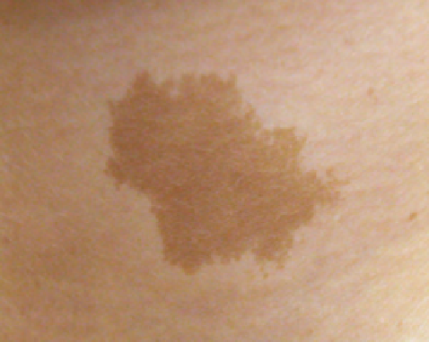
A 21-year-old woman with a history of large CALM on her left anterior neck presented for evaluation (Fig. 3.6A). Treatment
plan included monthly sessions of treatment with the alexandrite laser (755 nm). At the fi rst treatment session, the lesion was
treated with an 8-mm spot and a 50 J/cm
2
fl uence with multiple pulses. When she returned 6 weeks later, 50% clearance of the
lesion was noted. The lesion was again treated with an 8-mm spot, 60 J/cm
2
fl uence, and no cooling. When she returned
3 months later, the lesion was noted to be 90% cleared (Fig. 3.6B). The CALM was again treated with the alexandrite laser with
an 8-mm spot, a fl uence of 60 J/cm
2
, and 50/20 cryogen cooling with three treatments approximately 6 weeks apart to treat
the minimal amount of residual pigment.
(
A
)
(
B
)
Figure 3.6
Café-au-lait. (
A
) Before and (
B
) after six treatment sessions with the 755-nm alexandrite laser.
Nevus Spilus
Nevus spilus is a lesion in which darkly pigmented macules or
papules lie within a typical CALM. Estimated to occur in 2.3%
of patients (male and female equally) visiting a dermatology
practice, nevus spilus seems to be concentrated on the trunk
and lower extremities. Although nevus spilus is generally
not considered to be a precursor of malignant melanoma,
several studies have reported cutaneous melanomas within
these nevi.
Histologically, these lesions are composed of a lentiginous
elongation and hyperpigmentation of rete ridges with an
increase in melanocytes. Often a nesting of nevus cells occurs
within the lesion. The clinically dark speckled areas are either
junctional nevi or compound nevi. Although rare reports
of nevus spilus undergoing malignant degeneration suggest
exercising caution when treating them (31-34), many report
a good response to QS-ruby, QS-alexandrite, and QS-FD
532-nm Nd:YAG lasers (35,36). Unfortunately, some patients
respond poorly, and complete resolution may require four or
more treatments. In addition, the longer pulsed diode and
alexandrite lasers in the millisecond domain can be used on
the discrete nevi within the nevus spilus. The underlying
biology may explain the poor responders; not all the involved
melanocytes are targeted because of insuffi cient melanin. The
histology of these lesions reveals nothing that will further
explain those with a poor response. Treatment is the same as
for CALM.
laser treatment for
epidermal
-
dermal lesions
Becker's Nevus
Becker's nevi usually appear in childhood or early adult life as
a light- to medium-brown patch ranging in size from as small
as 2 cm to encompassing an entire shoulder. Lesions are