Biomedical Engineering Reference
In-Depth Information
Seborrheic Keratoses
Seborrheic keratoses are common benign epidermal prolifera-
tions that evolve from light-yellow, smooth macules to verrucous
pigmented papules or plaques. Although there are several histo-
pathologic variants of SK, the acantholytic type is the most com-
mon, consisting of interweaving bands of keratinocytes associated
with variable amounts of increased epidermal pigmentation.
Dermatosis papulosa nigra is an entity that is clinically and
histologically similar to SK and occurs in dark skin types. SK and
DPN respond similarly to the lasers discussed for lentigines, but
thicker lesions may require additional treatments (22-24). In
addition, for thicker lesions, use of the long pulsed alexandrite
laser is more effective and requires fewer treatments. However, it
is important to note laser therapy has not been shown to be any
more effective than treatment with non-laser destructive meth-
ods (cryotherapy and electrodessication) (27,28).
Case 5
Seborrheic Keratoses
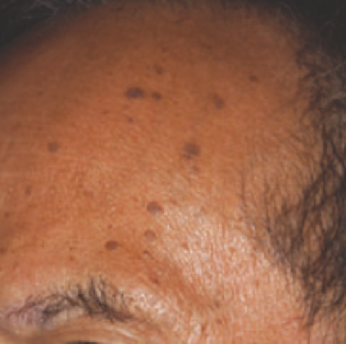
A 51-year-old man with a history of numerous SKs scattered over his face presented for treatment (Fig. 3.5A). Treatment
options were discussed, including liquid nitrogen, electrocautery, and laser treatment. Risks of possible pigmentary change
secondary to treatment were emphasized to the patient. A test spot was performed using the QS alexandrite laser at 6.5 J/cm
2
with a 3-mm spot size. When the patient returned 3 weeks later, the treated area was noted to have complete clearance without
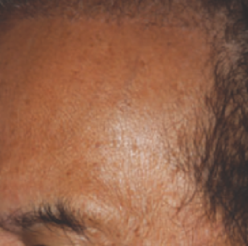
residual pigmentary change. A larger area over the left forehead was therefore treated at the same settings. Patient returned
6 weeks following treatment and was noted to have signifi cant improvement in his SKs (Fig. 3.5B).
(
A
)
(
B
)
Figure 3.5
Seborrheic keratoses (
A
) before and (
B
) 6 weeks after treatment with the QS alexandrite laser (755 nm).
Café-au-lait Macules
CALMs are light tan to brown hypermelanotic fl at lesions that
are sharply demarcated from the surrounding normal skin.
Found in up to 13.8% of the population, CALMs may appear
at birth or soon thereafter and increase in number over the
fi rst two decades of life. This lesion may be found in several
syndromes, including von Recklinghausen disease, Albright
syndrome, and Marfan syndrome.
Histologically, hypermelanosis is present in the basal
layer of the epidermis in melanocytes and keratinocytes. Giant
melanosomes, present in keratinocytes and basal melanocytes of
CALMs in patients with neurofi bromatosis, have not been
reported in other CALM patients. These lesions are not associ-
ated with malignant potential and are commonly treated for
cosmetic reasons. The QS ruby (5.0- to 6.5-mm spot size
with 5-8 J/cm
2
), QS alexandrite (3- to 5-mm spot size with
6-9 J/cm
2
), and FD QS Nd:YAG lasers (3- to 4-mm spot size with
1-4 J/cm
2
) have been used to treat CALMs (29,30). Longer
pulsed alexandrite lasers have been used in hopes of decreas-
ing the recurrence rate and also decreasing the number of treat-
ment sessions required. It is possible that the longer pulse width
allows for collateral thermal damage to non-pigment-containing