Biomedical Engineering Reference
In-Depth Information
(
A
)
(
B
)
Figure 1.36
Blood vessels (
A
) with and (
B
) without cross-polarization.
0.5-mm beam
1.0-mm beam
2.0-mm beam
(
A
)
(
B
)
(
C
)
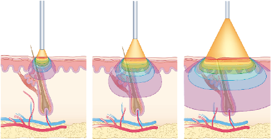
Figure 1.37
Note how spot size changes impact the depth of penetration. Shown are the fl uence patterns for 1064-nm light with beams (
A
) 0.5 mm, (
B
) 1.0 mm,
and (
C
) 2.0 mm in diameter on the skin surface.
references
1. Kono T, Isago T, Honda T, Nozaki M. Treatment of facial lentigines with
the long pulsed dye laser by compression method. Lasers Surg Med 2004;
34(Suppl 16): (abstract): 33.
2. Grossweiner L. The Science of Phototherapy. Boca Raton: CRC, 1994.
3. Hillenkamp F. Interaction between laser radiation and biological sys-
tems. In: Hillenkamp FRP, Sacchi C, eds. Lasers in Medicine and Biology.
Vol Series A New York: Plenum, 1980: 37-68.
4. Welch A, van Gemert MJ, Starr JC, Wilson BC. Defi nitions and over-
view of tissue optics. In: Welch AJ, van Gemert MJ, eds. Optical-Ther-
mal Response of Laser-Iradiated Tissue. New York: Plenum, 1995: 15-46.
5. Anderson RR, Parrish JA. The optics of human skin. J Invest Dermatol
1981; 77: 13-19.
6. van Gemert MJ, Jacques SL, Sterenborg HJ, Star WM. Skin optics. IEEE
Trans Biomed Eng 1989; 36: 1146-54.
after obvious removal of the epidermis, does not? Obviously,
much of the work in the future for scientists will involve a
more complete “mapping” of these complex responses.
Laser, nonlaser light sources, and other physical modalities
will undoubtedly play different roles than the traditionally
destructive ones that they have played to date. As we further
harness and understand EMR, the role of light will expand
into real-time bedside diagnostic tools. One day the derma-
tologist could well diagnose a BCC noninvasively, prepare the
patient for PDT, and months later “re-biopsy” the patient with
optical coherence tomography for test of cure! All of this
would occur without disruption of even the stratum corneum!
The future is indeed exciting.