Biomedical Engineering Reference
In-Depth Information
regions between tanned and untanned areas can be accounted
for (150). One newer device integrated into an IPL system is
the Skintel™, a built-in pigment meter that sends a Bluetooth
signal from the skin surface to the base device and suggests safe
start settings to the operator for that local anatomic region.
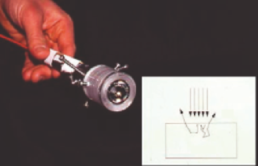
Using a Polarizing Lamp to Enhance Illumination
One can optimize laser treatment by using a polarizing lamp
during procedures to enhance the appearance of blood and
pigment dyschromias (Fig. 1.36A,B) (151).
Air
Selective Cell Targeting
A process called selected cell targeting has been examined as a
way to destroy selected cells. This precise energy deposition is
achieved by using laser pulses and light absorbing immunocon-
jugates tagged to the respective cells. The investigators in one
study showed, for example, that lymphocytes could be selec-
tively damaged by attaching iron oxide microparticles absorb-
ing 565-nm radiation at those sites (152). One can imagine, in
the future, using this type of modality to treat T-cell-mediated
diseases, such as atopic dermatitis or psoriasis. In this way, one
makes the “bad guy” more noticeable to the laser.
Tissue
Figure 1.35
Hemispheric mirror that “sits” over the skin and reflects remitted
photons back to the skin surface (so-called photon recycling).
1
RR
,
where
R
S
is the skin refl ectance
and
R
M
is that of a hemispherical mirror. For example, if
R
S
is
0.7 and
R
M
is 0.9, a gain of
skin is a factor of
(1
−
)
SM
1
0.63)
,
or almost threefold, can
(1
−
be achieved.
Scatter-Limited Therapy Using Small Microbeams
Reinisch (153) proposed the use of variously sized beams to
limit penetration into the dermis. By using the aforementioned
spot size arguments, one can exploit the properties of small spots
to change the way particular wavelengths behave in the skin. For
example, one can tailor a 1064-nm laser to heat progressively
larger depths of skin by increasing the spot size (Fig. 1.37).
Plasma for Skin Regeneration
Gas plasmas have been used in surgical devices for many years.
Typically, ionized gas acts as a conducting pathway to deliver RF
energy to achieve superfi cial tissue coagulation. The plasma
energy is relatively small, as the tissue effect is achieved primarily
by RF energy. In contrast, the Portrait PSR (plasma skin resurfac-
ing), (Rhytec, UK) uses ultrahigh-frequency (UHF) RF energy to
ionize a fl ow of nitrogen gas producing millisecond pulses of
plasma (3). The plasma, characterized by a lilac glow transition-
ing to a yellowish light called a Lewis-Rayleigh Afterglow as it
fl ows out of the nozzle of the handpiece, produces short-lived
rapid elevations in the temperature of the skin's surface. The dis-
ruptive effect of energy conversion through an intermediate
chromophore found with high-energy lasers is avoided. The
important clinical reason for selecting nitrogen is that it purges
oxygen from the skin's surface so that oxidative carbonization is
minimized, eliminating unpredictable “hot spots” and charring
that can produce scarring. A new pixilated RF plasma system (RF
Pixel, Alma Lasers, Buffalo Grove, IL, USA) uses a rolling set of
electrode tips to create microwounds in the skin (Fig. 1.29).
Two-Photon Excitation
We often are limited in dermatology by using VIS light laser
technologies with which are intrinsically nonpenetrating.
By using very high power density sources, there is a concept
known as “two-photon excitation” (154). This concept allows
for optimal beam penetration while still preserving tissue target
selectivity for shorter wavelengths. If, for example, one wants to
target a red tattoo deep in the skin with 532-nm light, high-
fl uence 1064-nm light can be applied to the surface. By chance
some photons will arrive simultaneously at the target, creating
one higher energy photon. Although presently only used in
diagnostic applications in the future, this sort of technology
might overcome some obstacles in our present approaches.
Enhanced Drug Delivery Through Microchannels
Investigators have examined the role for fractional lasers in drug
delivery through the skin. Already, enhanced delivery of ALA has
been reported (155,156). Another potential role for fractional
lasers is sequential fi ring whereby the fi rst pulse creates a gateway
for the second pulse. For example, a CO
2
laser vertical channel
could create a hole for a subsequent beam. The second beam
would have a “head start” and travels deeper into the skin (157).
Finally, the reader should note that most of this chapter has
dealt with the physical portion of the skin's reaction to light
and electricity. However, oftentimes a characterization of the
reaction between the tissue and the energy source is not as dif-
fi cult as the subsequent biological sequelae. For example, as
Dr. Martin van Gemert remarked, why do DE junction-derived
blisters (after KTP laser) often cause scars but CO
2
laser, even
Pigment/Erythema Meters to Assist in Guiding Parameters
Many laser complications stem from a failure of the operator
to consider subtle pigmentary differences between patients
and the transitory nature of pigmentary status in the same
patient. For example, a tanned Caucasian patient may appear
to be equally dark as an Asian-American patient who is
untanned. However, in LHR, the patient with the higher con-
stitutive coloration will normally show a greater epidermal
tolerance. Tanned skin is injured skin, and newly introduced
spectrophotometers can allow the operator to distinguish
between levels of coloration within the context of erythema
and time. Proper use of these handheld devices allows the
astute operator to adjust settings based on absolute pigment
levels as well as ratios of pigment to erythema. Also, step-off