Biomedical Engineering Reference
In-Depth Information
ablative laser systems
The use of pulsed or scanned CO
2
lasers for ablative skin resur-
facing (ASR) is a popular procedure for similar indications as
dermabrasion and chemical peels. The same principles of
thermal confi nement used in selective photothermolysis also
apply to minimizing the thermal injury from CO
2
laser vapor-
ization. In fair-skinned patients, the most common indication
for skin resurfacing is to treat chronic sun damage, wrinkles,
traumatic scars, surgical scars, and acne scars. In contrast, in
non-white-skinned patients, acne scarring is the most com-
mon indication for this procedure. Unfortunately, the risk of
prolonged or permanent dyspigmentation, especially PIH,
parallels the degree of the patient's constitutive skin color or
natural pigmentation: the darker the color, the greater the
potential (164-166).
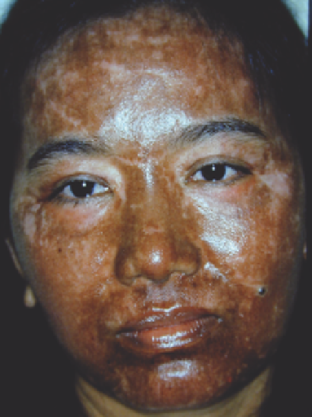
PIH, the most common complication seen following cuta-
neous CO
2
laser resurfacing in nonwhite patients, usually
develops around the fi rst month after treatment and becomes
most signifi cant within 4 months (Fig. 13.18). Various studies
reported an incidence of 25% and 68% on laser resurfacing in
Hispanic (skin phototypes II-V) (167) and other races with
skin type IV patients (168), respectively. This is compared with
a 3-7% incidence of PIH that occurs after CO
2
laser resurfac-
ing in Caucasian patients with skin phototypes I-IV. In fact, in
these studies, PIH occurred only in patients with skin photo-
types III and IV (164,165).
In contrast to PIH, which typically resolves with time, the
incidence of postlaser hypopigmentation is higher with a lon-
ger follow-up period. Incidence of PIH of 16-19% was noted
in an 8-month follow-up (164) and our 2-year follow-up
swelling. Laser tattoo removal on scar-prone areas, including
presternal, deltoid areas, and back should be performed with
the lowest fl uence possible.
As with treatment of all other pigmented lesions in pig-
mented races, the incidence of hypopigmentation appears to
be a wavelength-dependent phenomenon; the shorter the
wavelength, the greater the incidence of hypopigmentation.
The incidence of hyperpigmentation is comparable between
QSRL and QS Nd:YAG laser, which is mostly transient, and has
been reported only in darker-skinned patients (skin photo-
types II-V) (156,159). A dose-response study on the treat-
ment of tattoos by QSRL noted hypopigmentation at all doses
greater than 1.5 J/cm
2
. This persistent hypopigmentation was
apparent in four of 10 tattoos followed-up 1 year after treat-
ment (114). In contrast, hyperpigmentation was seen in only
one of 13 skin phototype V patients. The QSRL treatment
typically results in blistering at the dermoepidermal junction
(Fig. 13.4) (68), transient hypopigmentation and, less fre-
quently, hyperpigmentation (162).
As previously noted by others (154-156,159), we found that
when treating tattoos in Asian patients, the QSRL commonly
causes hypopigmentation, whereas the QS Nd:YAG laser at
appropriate fl uences has a very low incidence of hypopigmen-
tation. At the wavelength of 1064 nm, the QS Nd:YAG laser
light penetrates deeper and therefore might provide less injury
to the unintentionally targeted melanosomes (163). We there-
fore agree with the recommendation of others (151,154) that
the longer wavelength (1064 nm) QS Nd:YAG laser is prefera-
ble to the QSRL and QSAL in the treatment of deeper dermal
and blue/black tattoo pigments in dark-skinned patients.
(
A
)
(
B
)
Figure 13.18
(
A
) Melasma in a 40-year-old Thai woman with skin phototype IV, 2 weeks after test area resurfacing using an ultrapulsed CO
2
laser. No PIH was
seen. (
B
) Marked PIH developed 2 weeks after two passes of full-face CO
2
laser resurfacing.
Abbreviations
: CO
2
, carbon dioxide; PIH, postinfl ammatory hyperpig-
mentation.
Source
: From Ref. 168.