Biomedical Engineering Reference
In-Depth Information
(
A
)
(
B
)
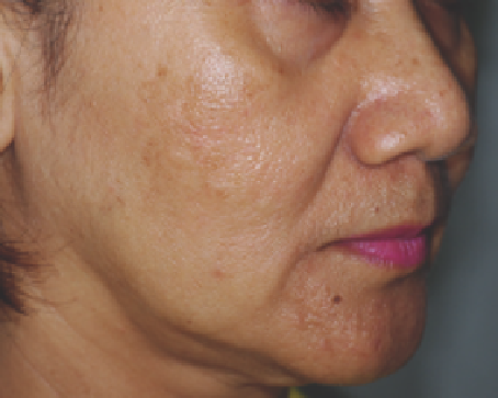
Figure 13.13
(
A
) A 50-year-old woman with melasma (skin type V) at baseline. (
B
) Clinical improvement 2 months after two variable square pulse erbium:yttrium-
aluminum-garnet laser treatments.
Source
: From Ref. 99.
The “threshold” radiant exposure response defi ned as imme-
diate whitening of pigmented lesions after short pulse laser
exposure is a useful clinical endpoint with any QS laser because
the whitening correlates directly with melanosome rupture
and pigment cell injury. Geronemus (111) treated nevus of
Ota in 15 patients of skin phototypes II-V with a QSRL and
noted that this threshold response was based on the patients'
skin phototype. Asian patients (skin types IV-V) required
slightly lower energy fl uence of 7.5-8.5 J/cm
2
to achieve imme-
diate whitening compared with white and Hispanic patients
(skin types II-IV) (8.5-10 J/cm
2
).
When treating nevus of Ota with QS lasers, lightening of the
lesion is noted after the fi rst session with additional clinical
improvement noted after every session. Multiple, sequential
treatments appear to increase the response rate and may be
required for complete clearing of the lesion (Fig. 13.14). The
response of nevus of Ota to QS laser treatment also appears to
depend on the color of the lesion. The maximum response rate
is found in the brown color, and gradually decreases in the
brown-violet, violet-blue, and blue-green colors, respectively.
On average, brown lesions can be cleared by three laser treat-
ments, brown-violet lesions by four sessions, violet-blue
lesions can be eliminated after fi ve sessions, and blue-green
lesions require at least six treatment sessions for complete
clearance (119).
Studies comparing the use of QSAL and QS Nd:YAG laser
showed that most patients more easily tolerated the former
(118). However, the QS Nd:YAG laser was found to be more
effective than the QSAL in the lightening of nevus of Ota after
three or more laser treatment sessions (76). In terms of adverse
effects, hypopigmentation is the most common adverse effect
after treatment with QS lasers (82) and can be permanent,
especially among those treated with QSRL (120). Transient
hyperpigmentation can be found especially after the fi rst treat-
ment session. The incidence of textural changes and scarring
are minimal.
Recurrence of original pigmentation could occur after com-
plete laser-induced clearing. The rate of recurrence is estimated
to be 0.6-1.2% (120). Laser treatment of nevus of Ota in chil-
dren demonstrated a better response and a lower risk of adverse
effects than in adults (121). Early treatment, leading to complete
clearance before school, may mean avoiding the childhood psy-
chological trauma associated with the cosmetic disfi gurement
of the birthmark. This advantage must now be weighed against
the risk of recurrence and the stress and cost associated with
multiple sessions of laser treatment.
In our current practice, patients are treated with a 1064-nm
QS Nd:YAG laser every 8-12 weeks. In a review of 125 patients
1-10 years postoperatively, we found that longer duration
between treatments provides a decrease in the total number of
treatments necessary over time (unpublished data, 2010). We
encourage the parents and/or the patients to start the treat-
ment as early as possible. Parents and patients are advised of
the risks of complications, including hyperpigmentation and
hypopigmentation, and are informed of the chance of recur-
rence after complete eradication. Ocular involvement includ-
ing elevated intraocular pressure with or without glaucoma
occurred in 10.3% of the patients; as a result, ophthalmologic
assessment is necessary (122).
Acquired Bilateral Nevus of Ota-like Macules (Hori's Nevus)
ABNOMs (Hori's nevus) is a common condition that affects
about 0.8% of the population with a marked female prepon-
derance (a male:female ratio of 1:6) (123). Clinically, ABNOMs
presents speckles or confl uent blue-brown or slate gray pig-
mentation that usually affects the bilateral malar areas. Other
involved sites include the temples, the root of the nose, the alae
nasi, the eye lids, and the forehead. Unlike in nevus of Ota, the
pigmentation in ABNOMs occurs in a symmetric bilateral
distribution, has a late onset in adulthood, and does not involve
mucosa. Histologically, lesions of ABNOMs demonstrate dif-
fuse upper dermal melanocytosis and differ from nevus of Ota,
in which dermal melanocytes occur not only in the upper der-
mis but also in the deep reticular dermis (124).
Pigment-specifi c lasers, including QSRL (125,126), QSAL
(127), and QS Nd:YAG (124,128-130) lasers have proved to be
effective in the treatment of ABNOMs. The treatment
responses have been noted to be less effective than those of the
nevus of Ota, and multiple sequential treatments are also
required to achieve the desired improvement (Fig. 13.5).