Biomedical Engineering Reference
In-Depth Information
(
A
)
(
B
)
(
C
)
(
D
)
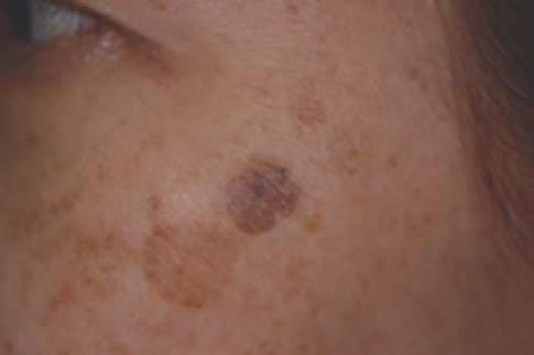
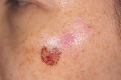
Figure 13.10
(
A
) A 60-year-old Thai woman with solar lentigo and fl at seborrheic keratosis on her cheek, before treatment. (
B
) An ash-gray color change without
purpura immediately after a treatment with a 595-nm PDL with compression at a fl uence of 9.0
J/cm
2
using a 7-mm spot size. (
C
) One week after treatment.
(
D
) Two weeks follow-up visit.
with compression, respectively. There was no signifi cant dif-
ference in the effi cacy between QSRL and PDL. However, com-
plications after PDL treatment were substantially less frequent
than after QSRL, presumably because of more optimal pulse
duration for targeting the basal cell layer, with minimal photo-
mechanical effect.
We achieved a similar degree of clearance when using this
PDL with compression technique to treat facial lentigines in
Thai and Chinese patients (Fig. 13.10). Lesions from various
anatomical regions were successfully treated, including back,
chest, upper/lower extremities, and face. We also treated a wide
range of lesions, including dark and light lentigines and fl at
seborrheic keratoses. Seborrheic keratoses and lighter lentigines
were more resistant to therapy. It is likely that the amount of
delivered procedures and percentage of more resistant lesions
in the patient group will dictate ultimate therapeutic outcome.
In addition, melasma tend to recur, often fairly soon, without
ongoing topical bleaching medications. In our current prac-
tice, sunscreen use is mandatory.
“Laser toning” using low-fl uence, large spot size, multi-
passed QS 1064-nm Nd:YAG laser for skin rejuvenation and
melasma has gained much popularity, especially in Asian
countries; however, there are still very few evidence-based data
supporting this approach in the treatment of PIH and melasma
(87-90). In laser toning, multiple passes of low-fl uence laser
(e.g., 1.6-3.5 J/cm
2
) are delivered through a large spot size
(e.g., 6-8 mm) to optimize energy delivery and to achieve mild
erythema as the clinical endpoint. Some physicians have pro-
posed the daily usage of laser toning for skin rejuvenation,
whereas others offer treatments at weekly, 2-weekly, or
monthly intervals with a wide variation in the total number of
treatments.
Wattanakrai et al. (89) noted statistically signifi cant improve-
ment of melasma from baseline in colorimeter and modifi ed
Melasma Area and Severity Index (mMASI) score after fi ve
laser-toning treatments in 22 Thai patients. However, 18% of
them developed rebound hyperpigmentation, and all had
recurrence of melasma at 3 months after the last laser session.
Although this treatment modality produced initial benefi cial
results to patients, it was not without adverse effects. During
the course of fi ve laser treatments, 14% of the patients experi-
enced faint, spotty hypopigmentation.
mixed epidermal and dermal
pigmented lesions
Postinflammatory Hyperpigmentation and Melasma
Mixed epidermal and dermal lesions, such as PIH or melasma,
respond variably and unpredictably to QS lasers (81-83) and
IPL (84-86). It is possible that not all pigment-producing struc-
tures are affected following a single irradiation. Therefore,
residing melanocytes may cause additional pigmentation in
patients tending to postlesional hyperpigmentation or melasma.