Biomedical Engineering Reference
In-Depth Information
In contrast, several studies by other investigators noted no
clinical improvement of striae and no increase in dermal elas-
tin content histologically (47,48). A study on the treatment of
striae in skin types IV and VI patients has demonstrated no
noticeable clinical improvement, with a higher risk of pigmen-
tary alterations (48). In our experience, we also found that
PDL provides a very minimal benefi cial effect on the treat-
ment of striae in Asian patients. Studies have demonstrated
the ability of nonablative (49), ablative (50), and fractional
lasers and radiofrequency device (51) to improve striae disten-
sae in Asian skin with minimal side effects.
In summary, treatment technique and parameters for
vascular-specifi c lasers in Asian skin are similar to those of
white skin (see chap. 2), but more care should be taken in
selecting an appropriate energy and in determining proper
treatment intervals. Because melanin acts as a competing
chromophore for vascular-specifi c lasers, to be effective the
starting energy density has to be higher than that used on fair
Caucasian skin. However, the improvement following this
procedure seems to be less effective and with a higher inci-
dence of epidermal damage than those seen in white-skinned
patients. Laser-induced pigmentary alterations should be
completely resolved before the delivery of additional laser
treatment, and to effect the optimal result from each session,
longer intervals may be necessary between laser treatments.
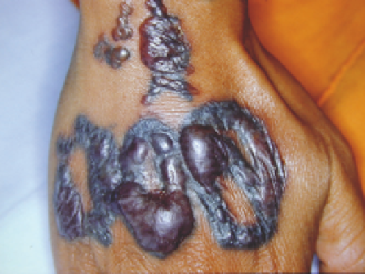
Figure 13.4
Blistering secondary to tattoo removal with the Q-switched ruby
laser in a skin phototype IV Thai patient using a fl uence of 6.5 J/cm
2
with a
5-mm spot size.
Source
: Courtesy of C. Vibhagool, Bangkok, Thailand.
with the argon [488 and 515 nm, continuous wave (CW)] laser
(54), 510-nm short PDL (55), copper vapor (511 nm, CW)
laser (56), QS frequency-doubled Nd:YAG (532 nm) laser
(57,58), QSRL (54,59-61), QSAL (755 nm) (62,63), the low-
fl uence carbon dioxide (CO
2
) (10,600 nm, CW) laser (64), and
IPL (65,66). All of these lasers and light sources carry a small
risk of depigmentation, hypopigmentation, and hyperpig-
mentation. However, when treating darkly pigmented skin,
pulsed lasers with appropriate energy density provide a more
selective destruction with a lower incidence of hyperpigmen-
tation and scarring as compared with continuous wave lasers
(5,67). The nonspecifi c injury to adjacent normal skin caused
from CW lasers may result in “laser tanning,” which has been
hypothesized to result from feedback inhibition of melano-
genesis, stimulation of tyrosinase activity, and/or release of
intracellular or extracellular melanocyte-stimulating factors
(68). This phenomenon is independent of PIH, which consti-
tutes another effect of the nonspecifi c damage caused from
CW laser energy.
pigment-specific lasers
Selective destruction of melanosomes has been well demon-
strated by exposing skin to submicrosecond, Q-switched (QS),
laser pulses (52,53). A wide range of these is available, including
a PDL (510 nm), QS frequency-double Nd:YAG laser (532 and
1064 nm), QSRL (694 nm), and QS alexandrite laser (QSAL)
(755 nm). All of these QS lasers are useful for treating superfi cial
epidermal lesions, such as lentigines and ephelides, café-au-lait
macules (CALMs), seborrheic keratosis, and dermal pigmented
lesions, such as blue nevus, nevus of Ota/Ito, ABNOMs (Hori's
nevus), infraorbital hyperpigmentation, drug-induced hyper-
pigmentaiton, and congenital melanocytic nevi.
Melanin absorption is stronger at shorter wavelengths,
whereas longer wavelengths penetrate better into the skin (18).
Several factors are involved in using QS lasers for treating benign
pigmented lesions in Asian individuals. First, the greater amount
of epidermal melanin results in greater damage to lesions and
adjacent normal skin pigment during laser irradiation. This
increased absorption may lead to posttreatment blistering
(Fig. 13.4), hyperpigmentation (Fig. 13.5), hypopigmentation
(Fig. 13.6), depigmentation, and even scarring. Second, larger
amounts of epidermal melanin in persons with dark skin tones
act as a competing chromophore for laser light while using these
QS lasers for treating dermal pigmented lesions. Thus, a larger
number of sequential treatments are required for complete
clearing compared with those of white-complexioned persons.
In addition, the adverse effects resulting from injury to epider-
mal melanin and the melanocytes responsible for producing
normal skin color should be anticipated.
Lentigines
Lentigines are a common sign of photoaging in Asians. This
epidermal pigmented lesion has shown excellent response to
QS (57) and LP Nd:YAG (532 nm) laser (69), QSRL (694 nm)
(60,61), QSAL (755 nm) (51), and low-fl uence CO
2
laser (52).
Our experience in treating Asian patients shows that the pulsed
dye (510 nm) laser, QS Nd:YAG laser (532 nm, 5-10 ns), QSRL,
and QSAL provide excellent results, usually with a single treat-
ment (Fig. 13.7).
The clinical endpoint is defi ned as the lowest fl uence that
can achieve immediate whitening (Fig. 13.8). This parameter
is about 2.0-2.5 J/cm
2
for the 532-nm QS Nd:YAG laser (10 ns,
3-mm spot diameter) (46), and about 7.0 J/cm
2
for the QSAL
(100-ns pulse width, 3-mm spot diameter) (62,66). For the LP
532-nm Nd:YAG laser the clinical endpoint, defi ned as a slate
gray appearance, is usually about 3.2 J/cm
2
(2- to 50-ms pulse
width, 2-mm spot diameter) (69).
QS (nanosecond domain) and LP (millisecond domain)
laser systems have been found to be equally effective in
Benign Epidermal Pigmented Lesions
Benign epidermal pigmented lesions include lentigines, ephe-
lides, CALMs, and seborrheic keratosis. In dark-skinned
patients, these pigmented lesions have been successfully treated