Biomedical Engineering Reference
In-Depth Information
(
A
)
(
B
)
(
C
)
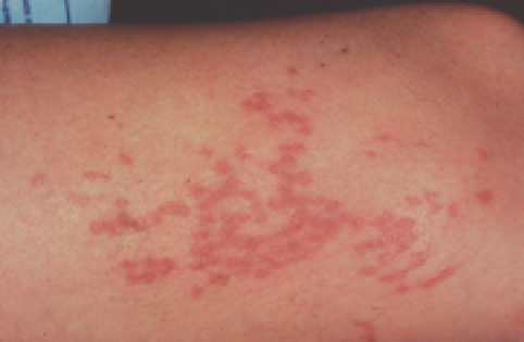
Figure 11.3
Photographic follow-up of telangiectatic fl are on the lateral thigh treated with fl ashlamp-pumped pulsed dye laser at 7 J/cm
2
, 125 pulses.
(
A
) Immediately before treatment. (
B
) Immediately after treatment; note the characteristic, localized urticarial response. (
C
) 6 weeks after treatment; note slight
hyperpigmentation and total resolution of telangiectasia. Pigmentation completely cleared over the subsequent 2-4 weeks.
Source
: From Ref. 30.
parameters were 30-ms pulses at 40 J/cm
2
. At these parameters,
vessels 0.4-1 mm in diameter showed 100% clearing in 22%,
75% clearing in 42%, and 50% clearing in 32%. Trelles et al.
(18) evaluated both the subjective and the objective effi cacy of
an 800-nm diode laser for leg vein clearance in 10 women
of various ages and skin types. Investigators used a sequence of
fi ve to eight stacked pulses with a pulse duration of 50 ms, a
delay of 50 ms, and a 3-mm spot size. Treatments were admin-
istered at 2-month intervals until complete clearance occurred,
and fi nal effi cacy was assessed 6 months following each
patient's fi nal treatment. To reach complete clearance, 50% of
patients needed three treatment sessions and the remaining
50% needed less than three. Although treated leg veins varied
from 1 to 4 mm in diameter, the best results were ultimately
seen in those vessels that had initially measured 3-4 mm.
Treatment of vessels located on the thigh as well as those in
patients with Fitzpatrick skin type III also yielded superior
effi cacy. Of note, no correlation was found between patient age
and effi cacy of treatment. A subsequent study evaluated the
dermal histologic and immunohistochemical changes induced
by exposure of leg telangiectasias to either a combination
915-nm diode laser and radiofrequency device or a 1064-nm
Nd:YAG laser (19). Among three patients having 0.1-2.0 mm
telangiectasias, each had one leg treated with the combination
diode and radiofrequency device and the opposite leg treated
with the Nd:YAG laser. Punch biopsies from treated areas were
taken 7 days after laser or radiofrequency exposure. Tissue
from each treatment type showed intermediate-sized vessels
with complete thrombosis and hemorrhage within the dermis
as well as the subcutis; focal full-thickness necrosis was seen in
the overlying epidermis. A single-treatment session of both
the combination diode and radiofrequency device and the
Nd:YAG, each yielded an average of 50-75% clinical clearing.
Thus, when comparing results from both treatment modali-
ties, a similar degree of improvement in the clinical appear-
ance of the telangiectasias was supported by histologic
examination of tissue specimens.
In summary, diode laser use is limited by treatment pain
and adverse effects. Of note, when feeding reticular veins
are not treated, distal treated telangiectasias tend to recur at
6-12 months after treatment. Some authors appear to be able
to achieve better results than others using similar parameters.
Although most apparent in target vessels larger than 1-2 mm
in diameter, the addition of radiofrequency to the diode
appears to yield vessel clearance at lower diode fl uences than
would be necessary to achieve the same results if the diode was
used alone.
high-intensity pulsed light
The high-IPL source was developed as an alternative to lasers
to maximize effi cacy in treating leg veins (Lumenis). This
device permits sequential rapid pulsing, longer duration
pulses, and penetrating longer wavelengths compared with
other laser systems. Theoretically, a phototherapy device that