Biomedical Engineering Reference
In-Depth Information
were seen in nine patients (47%), 1-6 months after the last
treatment. Keloids present for >2 years were more likely to
recur. Biopsies before and after treatment (6 months) docu-
mented a diminution or elimination of collagenous fi ber
whorls and of thick hyalinized collagen bands. Ki-67, a cell
proliferation marker, showed marked reduction (
p
= 0.0001).
Liver function tests, renal function tests, and CBC were nor-
mal before and after treatment.
Manuskiatti and Fitzpatrick (35) studied the response of
hypertrophic sternotomy scars to IL corticosteroids, IL 5-FU,
and fl ashlamp-pumped PDL treatments. They found all three
treatments to be roughly equivalent in effi cacy, but that corti-
costeroids had a much higher incidence of long-term sequelae
(atrophy, telangiectasia, and hypopigmentation), that is, 40%
versus 0%. The combined use of 5-FU with the V-beam Per-
fecta and the Fraxel Re:Store has become standard therapy for
scars in the practice of the senior author (Fig. 9.20).
Nanda and Reddy (189) reported a prospective uncontrolled
trial of IL 5-FU (50 mg/cc) given at weekly intervals for
12 weeks (0.5-2 cc per treatment) and then having follow-up
after an additional 24 weeks. Twenty-eight patients having
1-6 keloids were treated. Lesions ranged from 2 to 15 cm in
size and 6 months to 15 years in duration. Eight patients had
previously failed to respond to IL TAC 40 mg/cc every 3 weeks.
All patients responded to treatment, with 71% having good
response (>50% improvement) and 7% having excellent
response (>75% improvement). Flattening of the keloid and
peripheral regression were noted in all lesions. They noted no
difference in response of older keloids (15 years) and younger
lesions (6 months). Total and differential leukocyte counts
were normal at baseline, 12 weeks, and 24 weeks.
Asilian et al. (170) performed a 12-week single-blinded
clinical trial in which 69 patients with hypertrophic scars
or keloids were assigned to one of three groups. A single lesion
>10 mm in length was treated in each patient. All patients had
CBC, liver, and renal function tests at baseline and 12 weeks—
these were all normal. In Group 1, IL TAC (10 mg/cc) was
injected at weekly intervals for 8 weeks. In Group 2, TAC plus
5-FU (0.1 cc of 40 mg/cc TAC added to 0.9 cc of 5-FU 50 mg/cc)
was injected weekly for 8 weeks. Group 3 had TAC plus 5-FU as
in Group 2, but also added 585-nm pulsed dye laser (5-7.5 J/cm
2
)
at weeks 1, 4, and 8. Improvement was evaluated regarding
length, width, height of scars, erythema (visual scale), pliability
(5-point scale regarding induration), itching (4-point scale),
and patient self-assessment and overall improvement graded by
photos. Improvement in all areas was seen in each Group, but
TAC + 5-FU as well as TAC + 5-FU + PDL showed greater
improvement than TAC alone (
p
<0.05). Patient self-assessment
regarding >50% improvement was 20% in Group 1, 55% in
Group 2, and 75% in Group 3. Blinded observers rated >50%
improvement as 15% in Group 1, 40% in Group 2, 70% in
Group 3. Atrophy and telangiectasia were seen in 37% of those
treated with TAC alone, that is, Group 1.
Darougheh et al. (213) studied 40 patients with keloids in a
randomized double-blind clinical trial. Group 1 received TAC
10 mg/cc intralesionally, weekly for 8 weeks. Group 2 received
0.1 cc of TAC 4 mg mixed with 0.9 cc 5-FU 45 mg weekly for
8 weeks. Greater than 50% improvement at 12 weeks was
reported in 20% of Group 1 and 55% of Group 2, while trained
graders scored 15% in Group 1 and 40% of Group 2 as having
>50% improvement. Liver and renal function tests as well as
CBC were normal before and after 12 weeks.
(
A
)
(
B
)
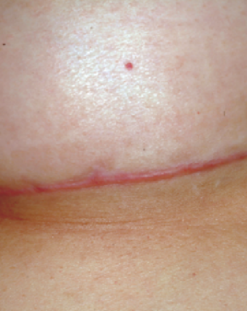
Figure 9.20
(
A
) Hypertrophic scar following breast augmentation. (
B
) Complete fl attening and softening of the scar has been achieved by the simultaneous use of
intralesional 5-FU, the V-beam Perfecta, and the Fraxel Re:Store.