Biomedical Engineering Reference
In-Depth Information
Plasma-Induced Ablation
With very high power densities exceeding 10
8
W/cm
2
, a phe-
nomenon called optical breakdown occurs. With plasma-
induced ablation, very clean and well-defi ned removal of
tissue without evidence of mechanical or thermal damage can
be achieved when choosing appropriate parameters. Plasmas
are sometimes produced by laser tattoo removal, where one
can observe a spark (19,27). Also, there is a new resurfacing
system that uses this fourth state of matter to target the skin
surface. The plasma is created by a radio frequency (RF)
excited N
2
gas, which is directed toward the skin. The plasma is
above the surface, creating very high but superfi cially confi ned
temperatures. The goal is to achieve damage to the skin surface
with minimal residual thermal damage (RTD). RF pixilated
devices can now create microplasmas on the skin surface to
correct acne scars and striae.
absorbing and scattering events. Characterization of the light
pathways is best understood by thinking of the incident beam
in terms of its constituent photons, where the photons statisti-
cally are either scattered or absorbed in a wavelength-dependent
fashion (43). The probabilities of absorption or scattering
(Table 1.1), designated
µ
a
and
µ
s
, respectively, are determined
by experiment. For a path length,
L
, the probability of photon
will not be absorbed or scattered is
e
−m
a
L
(2)
Jacques notes that a typical bloodless tissue value for
m
a
in the
VIS range is 1 cm
−1,
and the mean free path of a photon is
therefore 1 cm (44). For most VIS light, there are typically 100
scattering events before a photon is absorbed. As it turns out,
the photon scatters roughly 10 times before it loses its orienta-
tion with respect to the initial direction as it migrates in a ran-
dom walk. With scattering, there is
backscattered
light that
augments the delivered irradiance to yield a higher fl uence
beneath the tissue than at the tissue surface (Fig. 1.22) (44). An
often-used term is the penetration depth (
d
), which describes
the path length that causes 1/e attenuation of light. For a clear
solution,
d
accurately conveys the depth-dependent fl uence
skin optics
The optical properties of human skin determine the penetra-
tion, absorption, and internal dosimetry of laser light in skin.
The cosmetic surgeon can divide the skin into two main com-
ponents: (
i
) the epidermis (primarily an absorber of VIS light
due to melanin) and (
ii
) the dermis (which can be envisioned
as a carton of milk with red dots in it). When one uses a laser,
one should envision where the photons and/or electrical energy
is going and where the primary heating is. The laser surgeon
should memorize the absorption spectra of the main chromo-
phores in planning the procedure. He should remember that
the optical properties of the skin are not static. For example,
just a positional change in the arm will change the dermal
blood fraction. Also, just a few minutes in the sun will increase
the pigmentation index. Light-tissue interactions can be bro-
ken down into (
i
) the transport of light in tissue, (
ii
) absorption
of light and heat generation in tissue, (
iii
) localized tempera-
ture elevation in the target tissue (and denaturation of pro-
teins), and (
iv
) heat diffusion away from the target (Fig. 1.21).
The optical properties of the skin mimic those of a turbid
medium intermixed with focal discrete VIS and IR light
absorbers (blood, melanin, bilirubin, and dry collagen). There
is absorption by proteins, nucleic acids, and other compounds
in the UV spectrum, but outside of photochemistry, possibly
with a blue light source, these light-tissue interactions are
probably irrelevant for skin rejuvenation. In any light-tissue
interaction, the thermal or photochemical effects depend on
the
local
energy density at the target. Surface fl uence represents
the energy per unit area incident on the skin. Once the light
penetrates the surface, it undergoes a complex series of
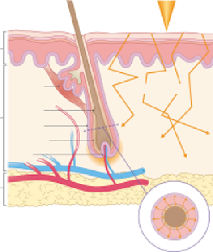
1. Laser
Pili
Muscle
2. Tissue optics
Bulge
Follicle
Melanin
Bulb
3. Heat source
4. Heat transfer
to follicle
Figure 1.21
The cascade of events in typical laser-tissue interaction with dis-
crete chromophore (in this case the hair follicle).
Table 1.1
Absorption Coeffi cients (cm
−1
) for Various Chromophores
Wavelength (nm)
410
532
595
694
755
810
940
1064
OxyHb (40% Hct)
1990
187
35
1.2
2.3
3.6
5.2
2.2
DeoxyHb
1296
138
96
6.6
5.2
2.7
3.0
0.6
Melanin
a
140
56
38
23
17
13
7
5.7
Water
6.7 × 10
−5
0.00044
0.0017
0.005
0.03
0.02
0.27
0.15
Bloodless dermis
10
3
2
1.2
0.8
0.6
0.5
0.4
OPD in skin (
μ
m)
100
350
550
750
1000
1200
1500
1700
a
Net epidermis for moderately pigmented adult: 10% melanin volume fraction in epidermis (46,141).
Abbreviations
: DeoxyHb, deoxyhemoglobin; Hct, hematocrit; OPD, optical penetration depth; OxyHb, oxyhemoglobin.