Biomedical Engineering Reference
In-Depth Information
3. Use feathering parameters to treat the eyelid
surfaces; use no more than two passes in the
immediate periorbital area except isolated spot
application to persisting folds of tissue.
4. Carefully observe the laser-tissue interaction of
the eyelids to avoid excessive tightening of tissue
and possible scleral show or ectropion, especially
in patients with a prior blepharoplasty.
5. Use the 3-mm beam to make a single pass along
the vermilion borders of the upper and lower lips,
because this is the most common area of persis-
tence of wrinkle lines. This also helps to emphasize
the vermilion border.
6. Do not routinely cross the vermilion border. Treat
up to the border, because this will make the lips
appear fuller and emphasize the vermilion border
as a result of the collagen tightening of the lip.
7. Treat lines that cross the vermilion border indi-
vidually by tracing over the line with the laser once
all the other areas have been treated, rather than
treating the entire vermilion surface.
8. After completion of treatment with the CPG,
carefully search the treatment surface for signs of
residual seborrheic keratoses, actinic keratoses, or
SCC in situ and for thickened scars, and vaporize
these to a fl at surface using the 3-mm beam with
pulse-stacking treatment technique or with the
Er:YAG laser.
9. Always carefully observe the immediate tissue
response regarding contraction or yellow-brown
discoloration. If this discoloration persists after
wiping with saline, this is a sign of thermal necro-
sis (Fig. 6.17).
10. Use the 3-mm spot in placing impact sites at 3- to
5-mm intervals to achieve tightening of the skin
without signifi cant thermal risk (concentric lines
of these treatment spots moving away from a site to
be tightened is effective). This is particularly useful
on the eyelids and midcheeks.
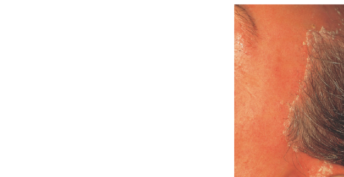
Figure 6.16
“Feathering” into hairline 5-15 mm to avoid leaving a line of con-
trast anterior to hairline. Hair will regrow normally in this feathered zone, but
it is not possible to treat the inside scalp without coagulating shaft of hairs.
The endpoint of treatment is when one of the following
conditions is seen:
1. The wrinkle or scar is removed.
2. A yellow-brown discoloration indicating thermal
damage is seen.
3. No further skin tightening is observed.
Figure 6.17
Persistent yellow-brown discoloration of dermis after wiping with
saline is a sign of thermal injury that may be extending deeper into dermis.
This skin-tightening effect allows achievement of compara-
ble clinical results at a more superfi cial level of tissue removal
than is necessary with other resurfacing modalities, such as
dermabrasion and chemical peels. The amount of tightening
observed depends on the number of passes delivered, unknown
specifi c tissue factors (possibly the numbers and health of
intact collagen fi bers), tissue thickness, and anatomic location.
Thin tissue such as eyelid and neck skin tightens very readily
and often profoundly, even to the point of causing ectropion
in eyelid skin. Temporal skin is generally thinner compared
with other facial skin and also tightens more signifi cantly.
Tightening of cheek skin may be important in reducing naso-
labial fold prominence and “jowls” under the jawline. Tighten-
ing may be used very selectively to achieve desired clinical
No reason exists to continue treating an area when any of
these signs is observed. Leave that area untreated from that
point on and concentrate on other tissue sites.
Collagen Shrinkage and Skin Tightening
It was not initially anticipated that collagen shrinkage would
be a component of the laser-tissue interaction. However,
unexpected improvement in redundant folds of the eyelids,
loose skin of the cheeks, nasolabial folds, and the other deep
wrinkle lines, coupled with the visible skin tightening
observable with the laser-tissue interaction, has led to the
use of heat-induced collagen shrinkage for clinical benefi t
(Box 6.1).