Biology Reference
In-Depth Information
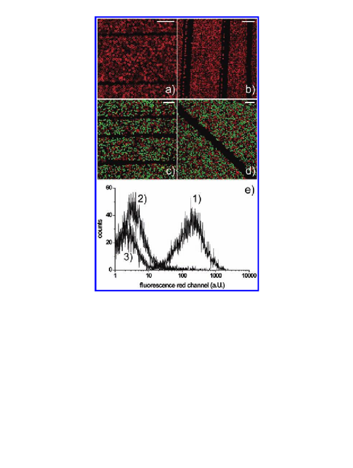
Figure 7.22
2D arrangement of composite LbL-lipid-virus colloids on slides
patterned with PAH. (a, b) Lipid-coated colloids arranged on a PAH-printed glass
slide. (c, d) Immunofluorescent assay on colloids fused with RV VLPs (green) as
negative control and Influenza A/PR8-coated colloids. The red signal indicates
specific binding of the primary antibodies as well as the secondary antibody on
colloids fused with Influenza A/PR8. Scale bars: 25
m. (e) Immunofluorescence
assay on colloids using flow cytometry. (e, assay 1) Colloids fused with Influenza A/
PR8 [geometric mean fluorescence (gmf), 163]. Controls: colloids fused with RLPs
(e, assay 2, gmf, 3.1), and lipid-coated colloids (e, assay 3, gmf, 2.5). Self-fluorescence
of lipid-coated colloids was 2.3 (data not shown). Reproduced with permission from
Fischlechner, M., Reibetanz, U., Zaulig, M., Enderlein, D., Romanova, J., Leporatti,
S., Moya, S., and Donath, E. (2007) Fusion of enveloped virus nanoparticles with
polyelectrolyte-supported lipid membranes for the design of bio/non-bio interfaces,
Nano Lett.
µ
,
7
(11), 3540-3546.
Search WWH ::

Custom Search