Biology Reference
In-Depth Information
Lvov
., 2005, 2006). Figure
7.20 depicts a multi-layered structure of CPMV particles and the polymer
pair linear poly(ethyleneimine) (PEI) and poly(acrylic acid) (PAA). CPMV
particles have a negative surface charge (under the conditions employed)
and bind stably to the positively charged PEI. An alternating structure of
polyelectrolyte with incorporated CPMV nanoparticles was self-assembled
(Steinmetz
et al
., 1994; Steinmetz
et al
., 2008b; Suci
et al
et al
., 2008b).
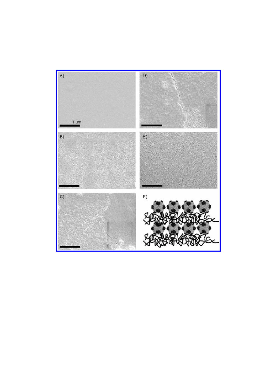
Figure 7.20
Scanning electron micrographs showing the sequential build-up of
polyelectrolytes and cowpea mosaic virus (CPMV) particles. (a) Precursor thin
film consisting of 2.5 bilayers of the polyions linear poly(ethyleneimine) (PEI) and
poly(acrylic acid) (PAA); (b) (PEI-PAA)
CPMV; (c) (PEI-PAA)
CPMV (PEIPAA); (d)
2.5
2.5
(PEI-PAA)
CPMV; (f ) schematic
representation of the architecture. All images were viewed at an acceleration voltage of
5 kV with a Zeiss Supra 55 VP FEG SEM; scale bar: 1
CPMV (PEI-PAA)
; (e) (PEI-PAA)
CPMV (PEI-PAA)
2.5
3
2.5
3
µ
m. Reproduced with permission
from Steinmetz, N. F., Findlay, K. C., Noel, T. R., Parker, R., Lomonossoff, G. P., and Evans,
D. J. (2008) Layer-by-layer assembly of viral nanoparticles and polyelectrolytes:
the film architecture is different for spheres versus rods,
ChemBioChem
,
9
(10),
1662-1670.
Search WWH ::

Custom Search