Biology Reference
In-Depth Information
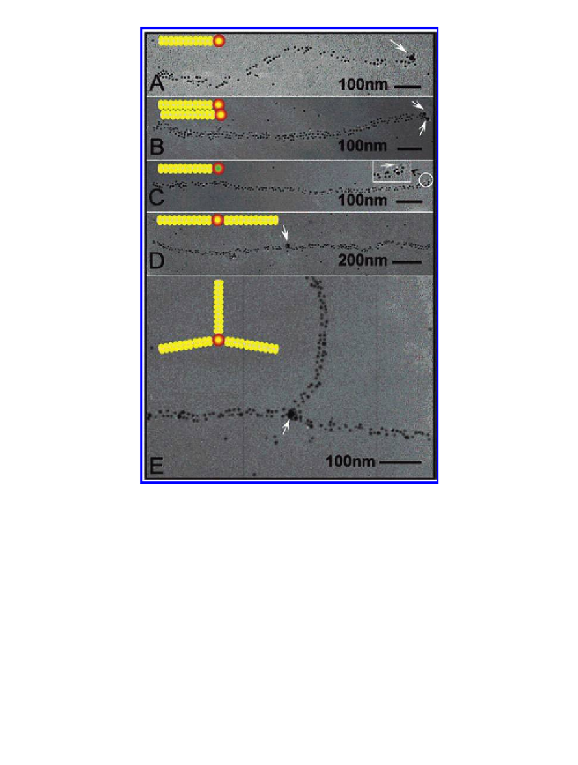
Figure 6.12
(a-e) Transmission electron micrographs of various nanoarchitectures
templated by M13. Gold nanoparticles (~5 nm) bind to genetically engineered
pVIII proteins along the virus axis and form 1D arrays, whereas a second peptide
motif on pIII protein simultaneously binds to streptavidin-coated nanoparticles.
Arrows highlight the streptavidin-conjugated gold nanoparticles (~15 nm) and CdSe
quantum dots bound on pIII proteins. The insets show the assembly schemes of
observed structures. White represents the virus structure, yellow dots represent gold
nanoparticles, the green dot represents a CdSe quantum dot, and red represents the
streptavidin coating around gold or CdSe particles. (c, inset) The enlarged image of
the CdSe quantum dot attached to the end of the virus. Reproduced with permission
from Huang, Y., Chiang, C. Y., Lee, S. K., Gao, Y., Hu, E. L., De Yoreo, J., and Belcher, A. M.
(2005) Programmable assembly of nanoarchitectures using genetically engineered
viruses,
Nano Lett.
,
5
(7), 1429-1434.
Search WWH ::

Custom Search